

Giant cell angioblastoma in an adult: a unique presentation
- PMID: 24179639
- PMCID: PMC3804802
- DOI: 10.4081/rt.2013.e27
Giant cell angioblastoma in an adult: a unique presentation
Abstract
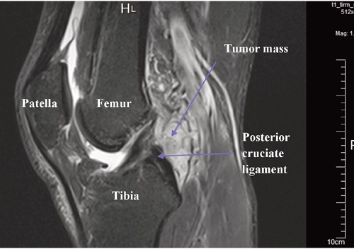
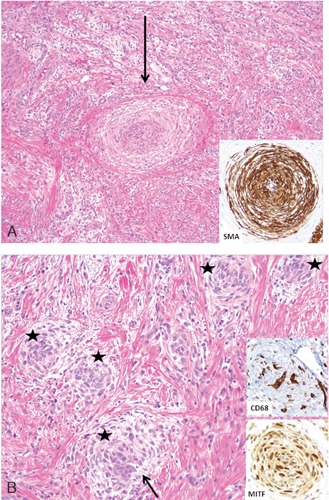
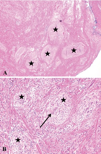
Giant cell angioblastoma is a very rare, locally destructive vascular tumor of intermediate malignancy without metastatic potential. There are only a few cases reported in the literature exclusively in the soft tissue of children. For the first time, we report on an adult patient with a giant cell angioblastoma in the popliteal fossa. The therapy included tumor resection with favorable clinical, oncological and functional outcome. Due to its locally destructive nature, surgery remains the mainstay of treatment. Histologically, giant cell angioblastoma is comprised of nodular aggregates of histiocytoid cells arranged around bland angiomatous spaces. Because of insufficient available data in regard to the definition of the entity, diagnostic criteria and its biological potential, it is not included in the new World Health Organization classification of tumors of soft tissue and bone. The differential diagnosis includes plexiform fibrohistiocytic tumor, myofibroma and giant cell fibroblastoma.
Keywords: angioblastoma; giant cell; intermediate malignancy; soft-tissue tumor; vascular tumor.
Figures
Similar articles
-
Giant cell angioblastoma of bone: four new cases provide further evidence of its distinct clinical and histopathological characteristics.Virchows Arch. 2015 Jul;467(1):95-103. doi: 10.1007/s00428-015-1757-0. Epub 2015 Mar 28. Virchows Arch. 2015. PMID: 25820372
-
Clinical manifestations and imaging and pathological features of giant cell angioblastoma: Report of four cases and literature review.Front Surg. 2023 Jan 5;9:1062309. doi: 10.3389/fsurg.2022.1062309. eCollection 2022. Front Surg. 2023. PMID: 36684227 Free PMC article. Review.
-
Giant cell angioblastoma: three additional occurrences of a distinct pathologic entity.Am J Surg Pathol. 2001 Feb;25(2):185-96. doi: 10.1097/00000478-200102000-00006. Am J Surg Pathol. 2001. PMID: 11176067
-
[Clinicopathologic study of giant cell angioblastoma].Zhonghua Bing Li Xue Za Zhi. 2010 Nov;39(11):752-6. Zhonghua Bing Li Xue Za Zhi. 2010. PMID: 21215166 Chinese.
-
Cutaneous and subcutaneous fibrohistiocytic tumors of intermediate malignancy: an update.Am J Dermatopathol. 2004 Apr;26(2):141-55. doi: 10.1097/00000372-200404000-00035. Am J Dermatopathol. 2004. PMID: 15024197 Review.
Cited by
-
Giant cell angioblastoma of bone: four new cases provide further evidence of its distinct clinical and histopathological characteristics.Virchows Arch. 2015 Jul;467(1):95-103. doi: 10.1007/s00428-015-1757-0. Epub 2015 Mar 28. Virchows Arch. 2015. PMID: 25820372
-
Clinical manifestations and imaging and pathological features of giant cell angioblastoma: Report of four cases and literature review.Front Surg. 2023 Jan 5;9:1062309. doi: 10.3389/fsurg.2022.1062309. eCollection 2022. Front Surg. 2023. PMID: 36684227 Free PMC article. Review.
References
-
- Goh SGN, Calonje E.Cutaneous vascular tumors: an update. Histopathology 2008;52:661-73 - PubMed
-
- Gonzalez-Crussi F, Cou P, Crawford SE.Congenital, infiltrating giant-cell angioblastoma. A new entity? Am J Surg Pathol 1991;15:175-83 - PubMed
-
- Mertens F, Unni K, Fletcher CDM.Pathology and genetics. Tumors of soft tissue and bone. Lyon: IARC Press; 2002
-
- Vargas SO, Perez-Atayde AR, Gonzalez-Crussi F, Kozakewich HP.Giant cell angioblastoma: three additional occurrences of a distinct pathologic entity. Am J Surg Pathol 2001;25:185-96 - PubMed
-
- Marler JJ, Rubin JB, Trede NS, et al. Successful antiangiogenic therapy of giant cell angioblastoma with interferon alfa 2b: report of 2 cases. Pediatrics 2002;109:E37. - PubMed
Publication types
LinkOut - more resources
Full Text Sources
Other Literature Sources
