

Reduced functional integration of the sensorimotor and language network in rolandic epilepsy
- PMID: 24179777
- PMCID: PMC3777786
- DOI: 10.1016/j.nicl.2013.01.004
Reduced functional integration of the sensorimotor and language network in rolandic epilepsy
Abstract
Introduction: Over the last years, evidence has accumulated that rolandic epilepsy (RE) is associated with serious cognitive comorbidities, including language impairment. However, the cerebral mechanism through which epileptiform activity in the rolandic (sensorimotor) areas may affect the language system is unknown. To investigate this, the connectivity between rolandic areas and regions involved in language processing is studied using functional MRI (fMRI).
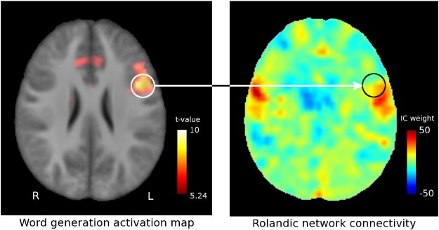
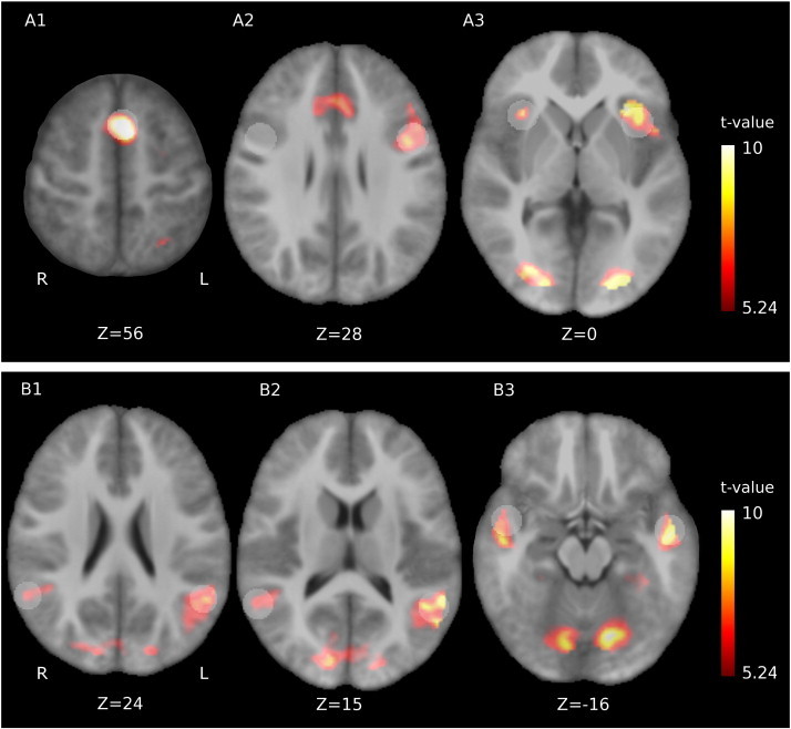
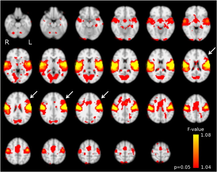
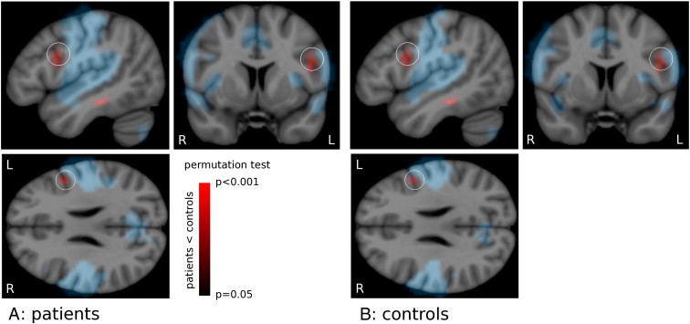
Materials and methods: fMRI data was acquired from 22 children with rolandic epilepsy and 22 age-matched controls (age range: 8-14 years), both at rest and using word-generation and reading tasks. Activation map analysis revealed no group differences (FWE-corrected, p < 0.05) and was therefore used to define regions of interest for pooled (patients and controls combined) language activation. Independent component analysis with dual regression was used to identify the sensorimotor resting-state network in all subjects. The associated functional connectivity maps were compared between groups at the regions of interest for language activation identified from the task data. In addition, neuropsychological language testing (Clinical Evaluation of Language Fundamentals, 4th edition) was performed.
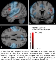
Results: Functional connectivity with the sensorimotor network was reduced in patients compared to controls (p = 0.011) in the left inferior frontal gyrus, i.e. Broca's area as identified by the word-generation task. No aberrant functional connectivity values were found in the other regions of interest, nor were any associations found between functional connectivity and language performance. Neuropsychological testing confirmed language impairment in patients relative to controls (reductions in core language score, p = 0.03; language content index, p = 0.01; receptive language index, p = 0.005).
Conclusion: Reduced functional connectivity was demonstrated between the sensorimotor network and the left inferior frontal gyrus (Broca's area) in children with RE, which might link epileptiform activity/seizures originating from the sensorimotor cortex to language impairment, and is in line with the identified neuropsychological profile of anterior language dysfunction.
Keywords: ICA, independent component analysis; Independent component analysis; Language impairment; RE, rolandic epilepsy; Resting-state fMRI; Resting-state networks; Rolandic epilepsy; Sensorimotor/rolandic network.
Figures
References
-
- Bak T.H., Chandran S. What wires together dies together: verbs, actions and neurodegeneration in motor neuron disease. Cortex. 2012;48:936–944. - PubMed
-
- Beckmann C.F., Mackay C.E., Filippini N., Smith S. OHBM annual meeting, San Francisco. 2009. Group comparison of resting-state FMRI data sing multi-subject ICA and dual regression.
-
- Berroya A.M., Bleasel A.F., Stevermuer T.L., Lawson J., Bye A.M. Spike morphology, location, and frequency in benign epilepsy with centrotemporal spikes. Journal of Child Neurology. 2005;20:188–194. - PubMed
LinkOut - more resources
Full Text Sources
Other Literature Sources
Miscellaneous
