

White matter microstructural abnormalities in bipolar disorder: A whole brain diffusion tensor imaging study
- PMID: 24179807
- PMCID: PMC3777761
- DOI: 10.1016/j.nicl.2013.03.016
White matter microstructural abnormalities in bipolar disorder: A whole brain diffusion tensor imaging study
Abstract
Background: Bipolar disorder (BD) is a chronic mental illness characterized by severe disruptions in mood and cognition. Diffusion tensor imaging (DTI) studies suggest that white matter (WM) tract abnormalities may contribute to the clinical hallmarks of the disorder. Using DTI and whole brain voxel-based analysis, we mapped the profile of WM anomalies in BD. All patients in our sample were euthymic and lithium free when scanned.
Methods: Diffusion-weighted and T1-weighted structural brain images were acquired from 23 lithium-free euthymic subjects with bipolar I disorder and 19 age- and sex-matched healthy control subjects on a 1.5 T MRI scanner. Scans were processed to provide measures of fractional anisotropy (FA) and mean and radial diffusivity (MD and RD) at each WM voxel, and processed scans were nonlinearly aligned to a customized brain imaging template for statistical group comparisons.
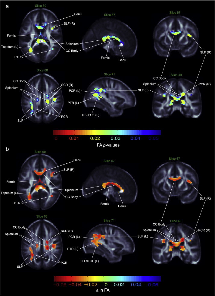
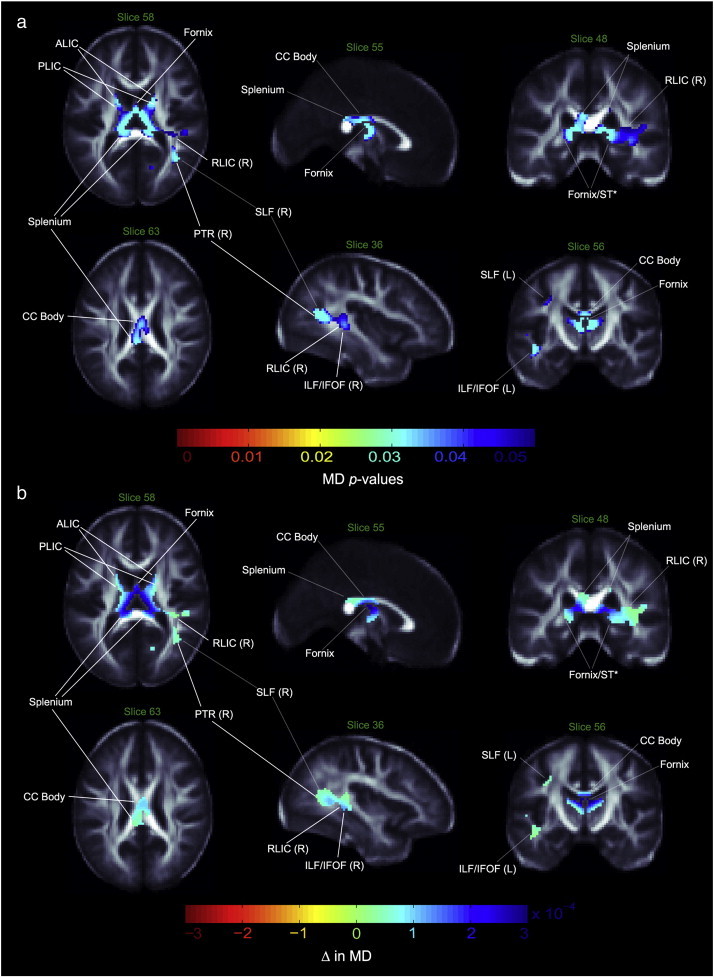
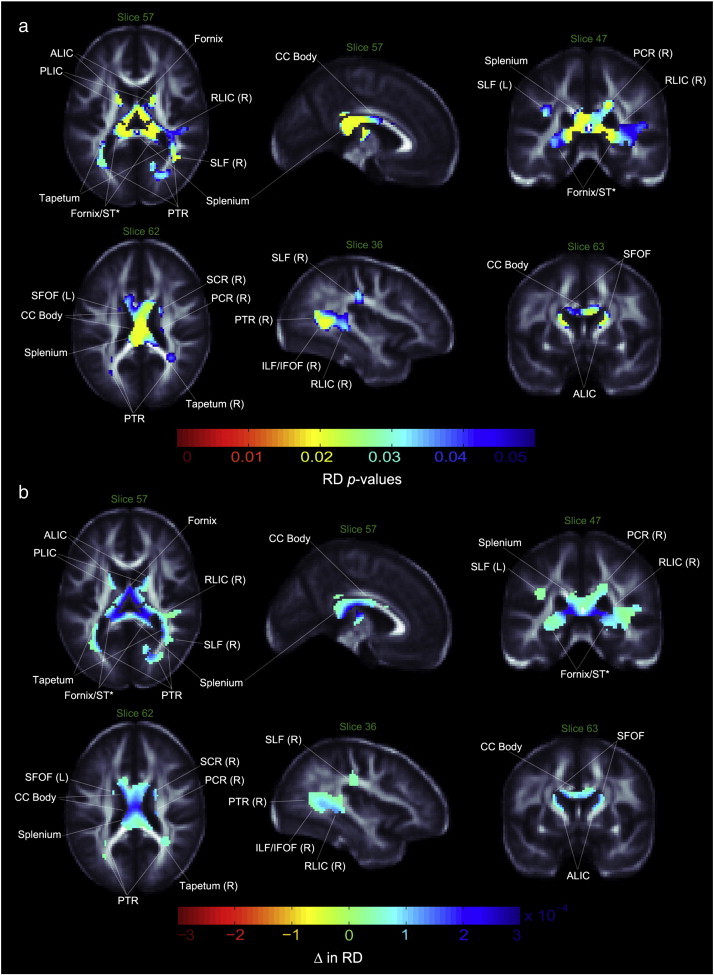
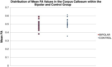
Results: Relative to controls, the bipolar group showed widespread regions of lower FA, including the corpus callosum, cortical and thalamic association fibers. MD and RD were abnormally elevated in patients in many of these same regions.
Conclusions: Our findings agree with prior reports of WM abnormalities in the corpus callosum and further link a bipolar diagnosis with structural abnormalities of the tapetum, fornix and stria terminalis. Future studies assessing the diagnostic specificity and prognostic implications of these abnormalities would be of interest.
Keywords: Bipolar disorder; Brain mapping; DTI; Fractional anisotropy; Neuroimaging; White matter.
Figures
References
-
- Adler C.M., Holland S.K., Schmithorst V., Wilke M., Weiss K.L., Pan H., Strakowski S.M. Abnormal frontal white matter tracts in bipolar disorder: a diffusion tensor imaging study. Bipolar Disorders. 2004;6:197–203. - PubMed
-
- Adler C.M., Adams J., DelBello M.P., Holland S.K., Schmithorst V., Levine A., Jarvis K., Strakowski S.M. Evidence of white matter pathology in bipolar disorder adolescents experiencing their first episode of mania: a diffusion tensor imaging study. The American Journal of Psychiatry. 2006;163:322–324. - PubMed
-
- Ahn M.S., Breeze J.L., Makris N., Kennedy D.N., Hodge S.M., Herbert M.R., Seidman L.J., Biederman J., Caviness V.S., Frazier J.A. Anatomic brain magnetic resonance imaging of the basal ganglia in pediatric bipolar disorder. Journal of Affective Disorders. 2007;104:147–154. - PubMed
-
- Altshuler L.L., Bookheimer S.Y., Townsend J., Proenza M.A., Eisenberger N., Sabb F., Mintz J., Cohen M.S. Blunted activation in orbitofrontal cortex during mania: a functional magnetic resonance imaging study. Biological Psychiatry. 2005;58:763–769. - PubMed
-
- FMRIB Analysis Group . The Oxford Centre for Functional Magnetic Resonance Imaging of the Brain, Department of Clinical Neurology, University of Oxford; Oxford, UK: 2012. FMRIB Software Library.
Grants and funding
LinkOut - more resources
Full Text Sources
Other Literature Sources
Miscellaneous
