. 2013 Nov 4:14:134.
doi: 10.1186/1471-2202-14-134.

Estrogen replacement modulates voltage-gated potassium channels in rat presympathetic paraventricular nucleus neurons

Affiliations

Estrogen replacement modulates voltage-gated potassium channels in rat presympathetic paraventricular nucleus neurons

Seul Ki Lee et al. BMC Neurosci. .

Abstract

Background: The hypothalamic paraventricular nucleus (PVN) is an important site in the regulation of the autonomic nervous system. Specifically, PVN neurons projecting to the rostral ventrolateral medulla (PVN-RVLM) play a regulatory role in the determination of the sympathetic outflow in the cardiovascular system. In the PVN-RVLM neurons, the estrogen receptor β is expressed. However, to date, the effects of estrogen on PVN-RVLM neurons have not been reported. The present study investigated estrogen-mediated modulation of two voltage-gated potassium channel (Kv) subunits, Kv4.2 and Kv4.3, that are expressed predominantly in PVN neurons and the functional current of Kv4.2 and Kv4.3, the transient outward potassium current (IA).

Results: Single-cell real-time RT-PCR analysis showed that 17β-estradiol (E2) replacement (once daily for 4 days) selectively down-regulated Kv4.2 mRNA levels in the PVN-RVLM neurons of ovariectomized female rats. There was no change in Kv4.3 levels. Whole-cell patch-clamp recordings demonstrated that E2 also diminished IA densities. Interestingly, these effects were most apparent in the dorsal cap parvocellular subdivision of the PVN. E2 also shortened a delay in the excitation of the PVN-RVLM neurons.

Conclusions: These findings demonstrate that E2 exerts an inhibitory effect on the functions of IA, potentially by selectively down-regulating Kv4.2 but not Kv4.3 in PVN-RVLM neurons distributed in a specific parvocellular subdivision.


Figures

Figure 1
Figure 1
Effects of E2 treatments on OVX rats. OVX rats were treated with vehicle (corn oil, 0.1 ml) and 17β-estradiol-3-benzoate (25 μg/kg in 0.1 ml corn oil) for the oil-group and the E2-group, respectively. A, the body weight-gain rate was compared between the two groups by calculating the ratio of body weight increase each day to the weight on the first day of treatment. A significant difference appeared on the third and the fourth days (*P < 0.05). B, the mean uterus weight was compared between the two groups. Uteri were isolated after sacrifice and weighed. The weight was normalized to the body weight on the day of sacrifice. E2 treatment significantly increased uterus weight (*P < 0.05).
Figure 2
Figure 2
Retrograde labeling of PVN-RVLM neurons. A, RVLM injection site was verified with serial medulla slices including RVLM (100 μm thickness). The representative image shows that a fluorescent spot was confined to the triangular frame defined as RVLM [29]. Amb, nucleus ambiguus; sp, the spinal trigeminal nucleus; py, pyramidal tracts; scale bar = 1 mm. B, the fluorescent image illustrates that the labeled PVN-RVLM neurons were located in the medial region (a) and in the posterior region (b) of the PVN in the hypothalamic brain slices (300 μm thickness). The PVN-RVLM neurons are evident primarily in the dorsal cap (DC), the ventral parvocellular (PaV), and the posterior parvocellular (PaPo) subdivisions. 3 V, the third ventricle; scale bar = 200 μm.
Figure 3
Figure 3
E2 effects on mRNA expression density of Kv channel subunits in single PVN-RVLN neurons. For relative quantification of Kv4.2 and Kv4.3 mRNA expression in the PVN-RVLM neurons between the oil- and the E2- groups, single-cell real-time RT-PCR was performed. A, the location of the PVN-RVLM neurons used for single-cell real-time RT-PCR in each subdivision. DC, the dorsal cap subdivision; PaV, the ventral parvocellular subdivision; PaPo, the posterior parvocellular subdivision. B, The mRNA expression density of the target genes of PVN-RVLM neurons was compared between the oil-group and the E2-group using the formula 2-ΔCt. C, the mRNA density of Kv4.2 and Kv4.3 was analyzed using the 2-ΔΔCt method and was finally expressed as n-fold that of the oil-group. The Kv4.2 significantly decreased, 0.54 ± 0.06 fold, in the E2-group (n = 21, *P < 0.05) compared to that of the oil-group (n = 19). However, Kv4.3 was not expressed differently between the two groups. The mRNA expression densities of Kv4.2 and Kv4.3 were compared in the three different subdivisions: the DC, the PaV, and the PaPo. In the DC, Kv4.2 mRNA expression significantly decreased in the E2-group, compared to that of the oil group, and decreased more dominantly than in overall expression. In the PaV and PaPo, no significant differences were detected. Kv4.3 mRNA expressions were not statistically different between the two groups for any subdivisions.
Figure 4
Figure 4
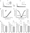
E2 effect on IA in PVN-RVLM neurons. A, representative images illustrate IA isolated in PVN-RVLM neurons from the oil-group (left) and the E2-group (right). The left inset indicates the voltage protocol used to isolate IA. The right inset indicates the reduction in IA by 4-aminopyridine (4-AP), the blocker of IA. B, Plots of IA current densities versus command step potentials. The current density was calculated by dividing the current amplitude of IA by cell capacitance. The E2 group (filled squares) displayed significant reductions in current densities of IA, compared to the oil-group (open squares). *P < 0.05. C, activation curve plotted with the mean normalized chord conductance (open squares for the oil-group and filled squares for the E2-group) and inactivation curve plotted with the mean normalized amplitude (open circles for the oil-group and filled circles for the E2-group). The curves were fitted with the Boltzmann equation. There were no significant differences in either the activation or the inactivation properties of IA. D, the maximal current density (at +25 mV step potential) was compared between the two groups. (a) In the E2-group, the maximal current density of IA significantly decreased, compared to that of the oil-group. When the differences between the two groups were compared according to subdivisions, a significant difference was detected only in the DC (b), not in either the PaV (c) or the PaPo (d). *P < 0.05. Insets in (b), (c) and (d) indicate the locations of the PVN-RVLM neurons used for IA recording in each subdivision. DC, the dorsal cap subdivision; PaV, the ventral parvocellular subdivision; PaPo, the posterior parvocellular subdivision.
Figure 5
Figure 5
E2 effects on delay of excitation in PVN-RVLM neurons. A, a representative image shows a delay in the onset of the first action potential expressed in PVN-RVLM neurons from the oil-group (dashed line) and the E2-group (solid line). The cells were hyperpolarized to near -90 mV to remove IA inactivation and depolarized by injecting a +45 pA current. B, The bar graph summarizes the differences in delay in the onset of the first action potential between the oil-group and the E2-group. The mean delay time of the E2-group significantly decreased, compared to that of the oil-group (*P < 0.05).

References

    1. Swanson LW, Kuypers HG. The paraventricular nucleus of the hypothalamus: cytoarchitectonic subdivisions and organization of projections to the pituitary, dorsal vagal complex, and spinal cord as demonstrated by retrograde fluorescence double-labeling methods. J Comp Neurol. 1980;194(3):555–570. doi: 10.1002/cne.901940306. - DOI - PubMed
    1. Pyner S, Coote JH. Identification of branching paraventricular neurons of the hypothalamus that project to the rostroventrolateral medulla and spinal cord. Neuroscience. 2000;100(3):549–556. doi: 10.1016/S0306-4522(00)00283-9. - DOI - PubMed
    1. Dampney RA. Functional organization of central pathways regulating the cardiovascular system. Physiol Rev. 1994;74(2):323–364. - PubMed
    1. Yang Z, Coote JH. Influence of the hypothalamic paraventricular nucleus on cardiovascular neurones in the rostral ventrolateral medulla of the rat. J Physiol. 1998;513(Pt 2):521–530. - PMC - PubMed
    1. Badoer E. Hypothalamic paraventricular nucleus and cardiovascular regulation. Clin Exp Pharmacol Physiol. 2001;28(1–2):95–99. - PubMed

Publication types

MeSH terms

Substances

LinkOut - more resources