. 2014 Jun;30(6):535-41.
doi: 10.1089/aid.2012.0195. Epub 2013 Nov 26.

Lymphocyte activation gene-3 expression defines a discrete subset of HIV-specific CD8+ T cells that is associated with lower viral load

Affiliations

Lymphocyte activation gene-3 expression defines a discrete subset of HIV-specific CD8+ T cells that is associated with lower viral load

José Peña et al. AIDS Res Hum Retroviruses. 2014 Jun.

Abstract

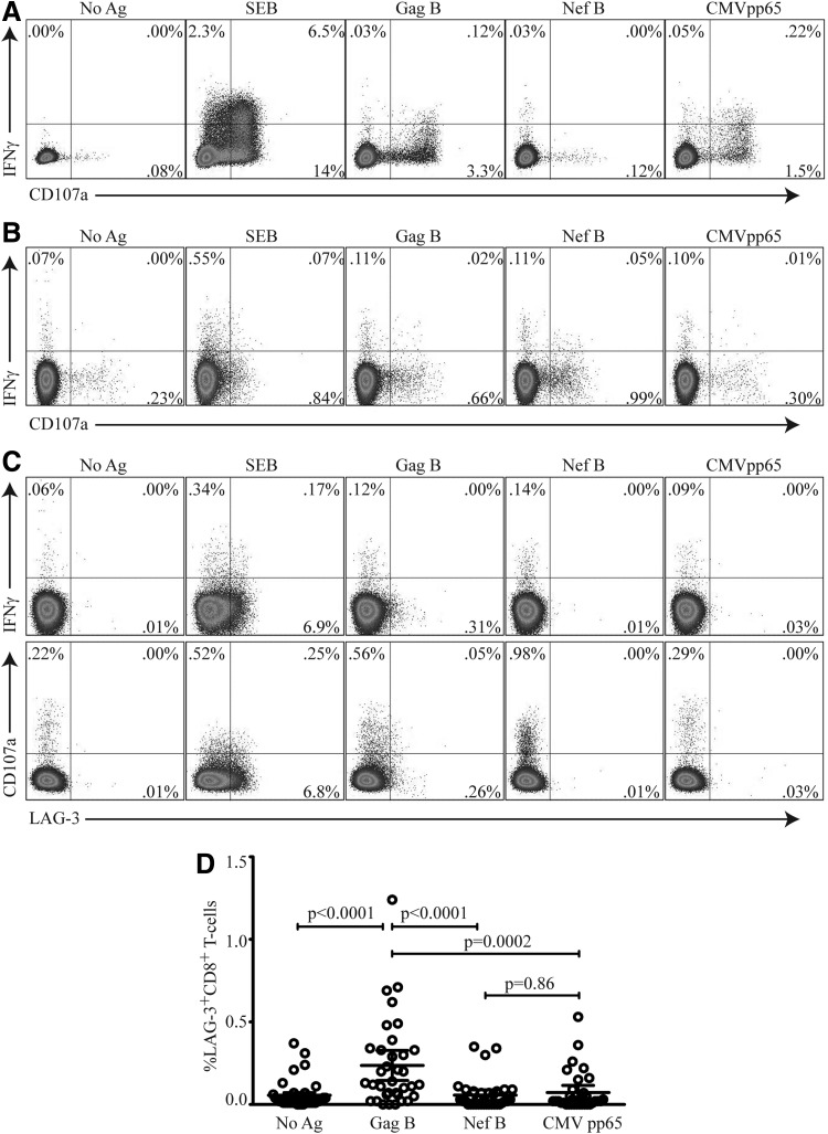
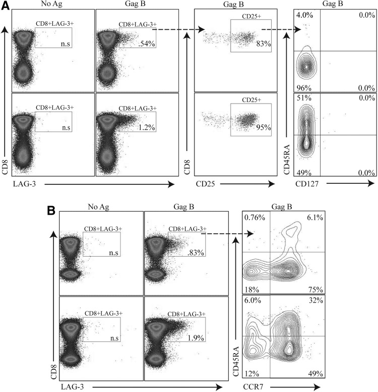
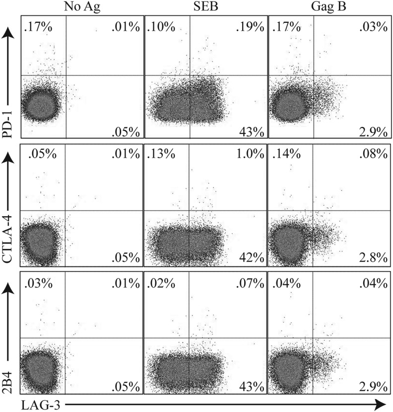
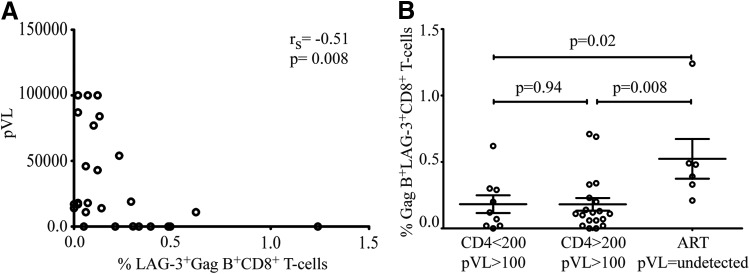
Mechanisms leading to the observed immune dysregulation in chronic HIV infection are not well understood. The MHC-II class ligand, lymphocyte activation gene-3 (LAG-3, CD223), has been implicated in the complex regulation mechanism of immune functions. In this study, we describe a new population of HIV-specific CD8(+) T cells expressing LAG-3. These LAG-3(+)CD8(+) T cells do not display immunophenotypic patterns traditionally attributed to regulatory T cells. The LAG3(+)CD8(+) T cells are CCR7(+),CD127(-), and display heterogeneous surface expressions of CD45RA and CD25. Interestingly, HIV-specific LAG-3(+)CD8(+) T cells do not substantially express CTLA-4 and LAG-3 expression does not correlate with interleukin (IL)-10 or tumor growth factor (TGF)-β production. In addition, HIV-specific LAG3(+)CD8(+) T cells do not produce interferon (IFN-γ) or express CD107a. The frequency of HIV-specific LAG3(+)CD8(+) T cells negative correlated with plasma viral load. Our study introduces a new population of HIV-specific CD8(+) T cells and proposes additional mechanisms of immune regulation in chronic HIV infection.


Figures

<b>FIG. 1.</b>
FIG. 1.
Lymphocyte activation gene-3 (LAG-3+) expression on HIV-specific CD8+ T cells does not express CD107a or produce interferon (IFN)-γ. (A) Peripheral blood mononuclear cells (PBMCs) were stimulated for 6 h with HIV antigens Gag B, Nef B, or CMV pp65 in the presence of anti-CD107a PE and Golgi-Stop or (B) restimulated following an initial 24-h stimulation, as described in Materials and Methods, then stained with Vivid viability dye for dead/live cell exclusion, anti-CD3 AmCyan, anti-CD4 PerCP Cy5.5, anti-CD8 APC-Cy7, anti-LAG-3 FITC, and IFN-γ APC, and analyzed by flow cytometry. Samples were gated on the CD3+/CD8+ lymphocyte population and then the percent of CD107a and IFN-γ-positive CD8+ T cells was determined. (C) Representative plots of the frequency LAG-3 on Gag- or Nef-specific CD8+ T cells and CMV pp65-specific CD8+ T cells expressing CD107a and IFN-γ are shown. Staphylococcus enterotoxin B (SEB) is used as a positive control. No significant overlap between Lag-3 expression and CD107a or IFN-γ was observed. (D) LAG-3+CD8+ T cell expression after antigen peptide stimulation compared to no antigen-stimulated control. Gag B-specific LAG-3+CD8+ T cells were statistically significant compared to Nef B or CMV pp65 (p<0.0001 and p=0.0002, respectively). Results were expressed as the percentage of CD107a or IFN-γ-positive CD8+ T cells (percent positive=percent Ag-specific – percent negative control). Responses ≥0.1% and two times the background were considered positive. p values <0.05 were considered significant (n=35).
<b>FIG. 2.</b>
FIG. 2.
HIV-specific LAG-3+CD8+ T cells express a variable surface immunophenotype. PBMCs were stimulated with HIV peptides then stained with Vivid for live/dead cell exclusion, LAG-3 FITC, CTLA-4 PE-Cy5, CD3 AmCyan, CD8 APC H7, CD25 PE, CD127 Alexa Fluor 647, and CD45RA PE-Cy7 and analyzed by flow cytometry. Samples were first gated on SSC/FSC, followed by SSC/CD3+, and then on the CD3+/CD8+ lymphocyte population; the percent of LAG-3+CD8+ T cells was then determined. (A) Two representative plots of HIV-specific LAG-3+CD8+ T cells after Gag B peptide stimulations that express CD25+ and are CD127 low. (B) HIV-specific LAG-3+CD8+ T cells with variable expression of CD45RA and CCR7. Box inserts within each quadrant represent the indicated cell subpopulations. Dotted arrows indicate further subgating of indicated cell subpopulations.
<b>FIG. 3.</b>
FIG. 3.
LAG-3+CD8+ T cell populations are distinct from PD1+, CTLA4+, or 2B4+ CD8+ T cells. PBMCs were stimulated with HIV-specific peptide Gag B peptides then stained with Vivid for live/dead cell exclusion, LAG-3 FITC, 2B4 PE, CD3 AmCyan, PD-1 APC, CTLA-4 PE-Cy5, CD4 PE-Cy7, and CD8 APC-H7. Samples were first gated on the CD3+/CD8+ lymphocyte population; the percent of LAG3+, PD-1+ CTLA4+, or 2B4+ cells was then determined. Representative plots of the frequencies of the LAG-3+ Gag-specific CD8+ T cells and expression of PD-1+ CTLA4+ or 2B4+ are shown. Responses ≥0.1% and two times the background were considered positive.
<b>FIG. 4.</b>
FIG. 4.
Negative correlation between the frequency of HIV-specific LAG-3+CD8+ T cells and HIV plasma RNA. The percentage of Gag-specific LAG-3 is plotted against the HIV plasma RNA. A significant negative correlation between HIV plasma RNA and Gag-specific LAG-3+CD8+ T cells in (A) was observed (rs=−0.51, p=0.0008). (B) Increased LAG-3 expression is also observed in antiretroviral treatment (ART)-mediated viral suppression compared to samples from individuals with CD4 <200 plasma viral load (pVL) >100 and CD4 >200 pVL >100 (p=0.02 and p=0.008, respectively); CD4 >200 (n=19), CD4 <200 (n=9), ART (n=5). Spearman rank correlations with p values<0.05 were considered significant.

References

    1. Triebel F, Jitsukawa S, Baixeras E, et al. : LAG-3, a novel lymphocyte activation gene closely related to CD4. J Exp Med 1990;171:1393–1405 - PMC - PubMed
    1. Workman CJ, Rice DS, Dugger KJ, Kurschner C, and Vignali DAA: Phenotypic analysis of the murine CD4-related glycoprotein, CD223 (LAG-3). Eur J Immunol 2002;32:2255–2263 - PubMed
    1. Workman CJ, Dugger KJ, and Vignali DA: Cutting edge: Molecular analysis of the negative regulatory function of lymphocyte activation gene-3. J Immunol 2002;169:5392–5395 - PubMed
    1. Baixeras E, Huard B, Miossec C, et al. : Characterization of the lymphocyte activation gene 3-encoded protein. A new ligand for human leukocyte antigen class II antigens. J Exp Med 1992;176:327–337 - PMC - PubMed
    1. Kisielow M, Kisielow J, Capoferri-Sollami G, and Karjalainen K: Expression of lymphocyte activation gene 3 (LAG-3) on B cells is induced by T cells. Eur J Immunol 2005;35:2081–2088 - PubMed

Publication types

MeSH terms