

Integrated imaging instrument for self-calibrated fluorescence protein microarrays
- PMID: 24182114
- PMCID: PMC3799691
- DOI: 10.1063/1.4823790
Integrated imaging instrument for self-calibrated fluorescence protein microarrays
Abstract
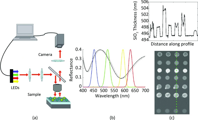
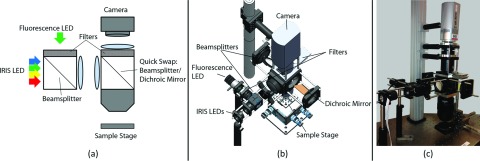
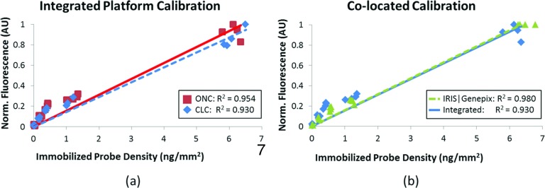
Protein microarrays, or multiplexed and high-throughput assays, monitor multiple protein binding events to facilitate the understanding of disease progression and cell physiology. Fluorescence imaging is a popular method to detect proteins captured by immobilized probes with high sensitivity and specificity. Reliability of fluorescence assays depends on achieving minimal inter- and intra-assay probe immobilization variation, an ongoing challenge for protein microarrays. Therefore, it is desirable to establish a label-free method to quantify the probe density prior to target incubation to calibrate the fluorescence readout. Previously, a silicon oxide on silicon chip design was introduced to enhance the fluorescence signal and enable interferometric imaging to self-calibrate the signal with the immobilized probe density. In this paper, an integrated interferometric reflectance imaging sensor and wide-field fluorescence instrument is introduced for sensitive and calibrated microarray measurements. This platform is able to analyze a 2.5 mm × 3.4 mm area, or 200 spots (100 μm diameter with 200 μm pitch), in a single field-of-view.
Figures
Similar articles
-
Label-Free and High-Throughput Detection of Biomolecular Interactions Using a Flatbed Scanner Biosensor.ACS Sens. 2017 Oct 27;2(10):1424-1429. doi: 10.1021/acssensors.7b00263. Epub 2017 Oct 4. ACS Sens. 2017. PMID: 28929734
-
Interferometric silicon biochips for label and label-free DNA and protein microarrays.Proteomics. 2012 Oct;12(19-20):2963-77. doi: 10.1002/pmic.201200202. Epub 2012 Sep 24. Proteomics. 2012. PMID: 22930463 Review.
-
Instrument-Free Protein Microarray Fabrication for Accurate Affinity Measurements.Biosensors (Basel). 2020 Oct 29;10(11):158. doi: 10.3390/bios10110158. Biosensors (Basel). 2020. PMID: 33138051 Free PMC article.
-
Quantitative characterization of conformational-specific protein-DNA binding using a dual-spectral interferometric imaging biosensor.Nanoscale. 2016 Mar 14;8(10):5587-98. doi: 10.1039/c5nr06785e. Nanoscale. 2016. PMID: 26890964
-
Diagnostic microarrays in hematologic oncology: applications of high- and low-density arrays.Mol Diagn Ther. 2009;13(2):91-102. doi: 10.1007/BF03256318. Mol Diagn Ther. 2009. PMID: 19537844 Review.
Cited by
-
Interferometric Reflectance Imaging Sensor (IRIS)--A Platform Technology for Multiplexed Diagnostics and Digital Detection.Sensors (Basel). 2015 Jul 20;15(7):17649-65. doi: 10.3390/s150717649. Sensors (Basel). 2015. PMID: 26205273 Free PMC article. Review.
-
Highly-Sensitive, Label-Free Detection of Microorganisms and Viruses via Interferometric Reflectance Imaging Sensor.Micromachines (Basel). 2023 Jan 21;14(2):281. doi: 10.3390/mi14020281. Micromachines (Basel). 2023. PMID: 36837980 Free PMC article. Review.
-
A Reliable, Label Free Quality Control Method for the Production of DNA Microarrays with Clinical Applications.Polymers (Basel). 2021 Jan 21;13(3):340. doi: 10.3390/polym13030340. Polymers (Basel). 2021. PMID: 33494542 Free PMC article.
References
-
- Kassahn K. S., J. Fish Biol. 72, 2407 (2008).10.1111/j.1095-8649.2008.01890.x - DOI
Publication types
MeSH terms
Grants and funding
LinkOut - more resources
Full Text Sources
Other Literature Sources