Three-dimensional analysis of maxillary changes associated with facemask and rapid maxillary expansion compared with bone anchored maxillary protraction
- PMID: 24182587
- PMCID: PMC3972125
- DOI: 10.1016/j.ajodo.2013.07.011
Three-dimensional analysis of maxillary changes associated with facemask and rapid maxillary expansion compared with bone anchored maxillary protraction
Abstract
Introduction: Our objectives in this study were to evaluate in 3 dimensions the growth and treatment effects on the midface and the maxillary dentition produced by facemask therapy in association with rapid maxillary expansion (RME/FM) compared with bone-anchored maxillary protraction (BAMP).
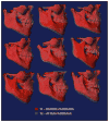
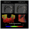
Methods: Forty-six patients with Class III malocclusion were treated with either RME/FM (n = 21) or BAMP (n = 25). Three-dimensional models generated from cone-beam computed tomographic scans, taken before and after approximately 1 year of treatment, were registered on the anterior cranial base and measured using color-coded maps and semitransparent overlays.
Results: The skeletal changes in the maxilla and the right and left zygomas were on average 2.6 mm in the RME/FM group and 3.7 mm in the BAMP group; these were different statistically. Seven RME/FM patients and 4 BAMP patients had a predominantly vertical displacement of the maxilla. The dental changes at the maxillary incisors were on average 3.2 mm in the RME/FM group and 4.3 mm in the BAMP group. Ten RME/FM patients had greater dental compensations than skeletal changes.
Conclusions: This 3-dimensional study shows that orthopedic changes can be obtained with both RME/FM and BAMP treatments, with protraction of the maxilla and the zygomas. Approximately half of the RME/FM patients had greater dental than skeletal changes, and a third of the RME/FM compared with 17% of the BAMP patients had a predominantly vertical maxillary displacement.
Copyright © 2013 American Association of Orthodontists. Published by Mosby, Inc. All rights reserved.
Conflict of interest statement
All authors have completed and submitted the ICMJE Form for Disclosure of Potential Conflicts of Interest, and none were reported.
Figures


References
-
- Turley PK. Orthopedic correction of Class III malocclusion with palatal expansion and custom protraction headgear. J Clin Orthod. 1988;22:314–25. - PubMed
-
- Hickam JH. Maxillary protraction therapy: diagnosis and treatment. J Clin Orthod. 1991;25:102–13. - PubMed
-
- Baik HS. Clinical results of maxillary protraction in Korean children. Am J Orthod Dentofacial Orthop. 1995;108:583–92. - PubMed
-
- Ngan PW, Hagg U, Yiu C, Wei SH. Treatment response and long-term dentofacial adaptations to maxillary expansion and protraction. Semin Orthod. 1997;3:255–64. - PubMed
-
- Macdonald KE, Kapust AJ, Turley PK. Cephalometric changes after the correction of Class III malocclusion with maxillary expansion/facemask therapy. Am J Orthod Dentofacial Orthop. 1999;116:13–24. - PubMed
Publication types
MeSH terms
Grants and funding
LinkOut - more resources
Full Text Sources
Other Literature Sources

