

Midpalatal suture maturation: classification method for individual assessment before rapid maxillary expansion
- PMID: 24182592
- PMCID: PMC4185298
- DOI: 10.1016/j.ajodo.2013.04.022
Midpalatal suture maturation: classification method for individual assessment before rapid maxillary expansion
Abstract
Introduction: In this study, we present a novel classification method for individual assessment of midpalatal suture morphology.
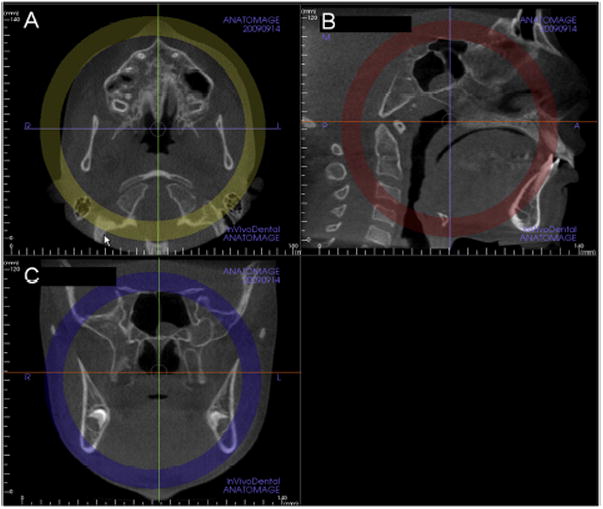
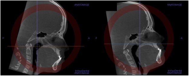
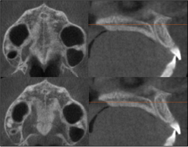
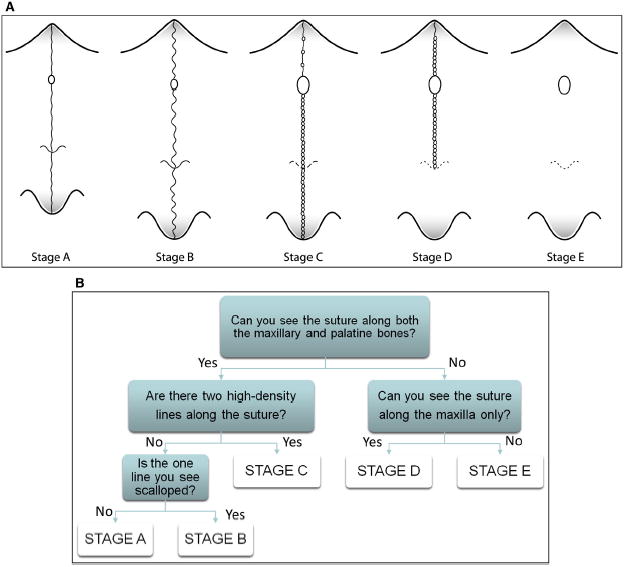
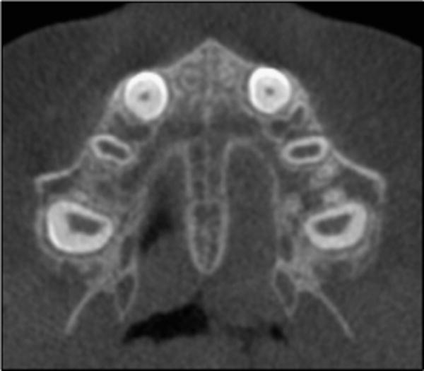
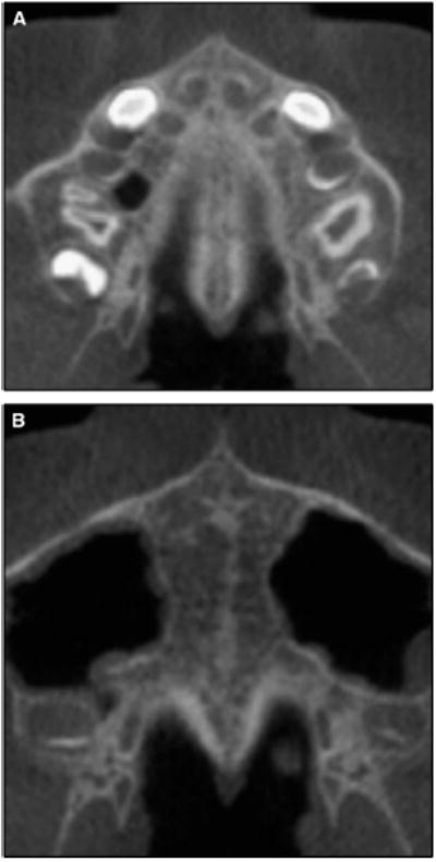
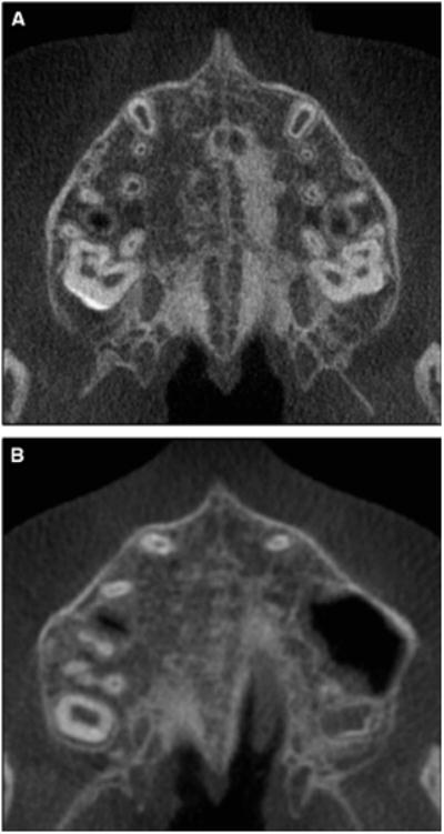
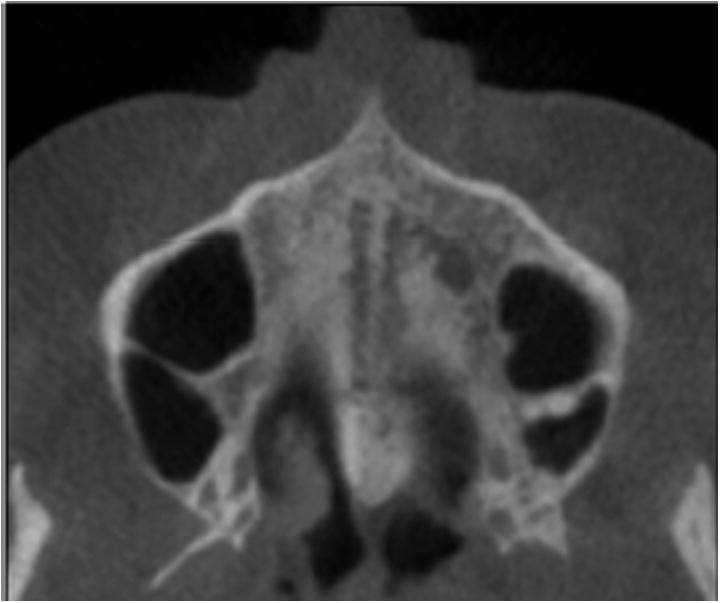
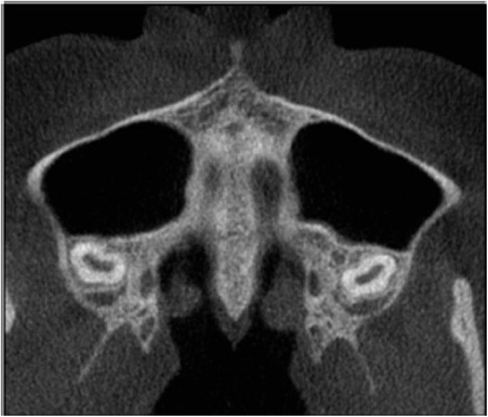
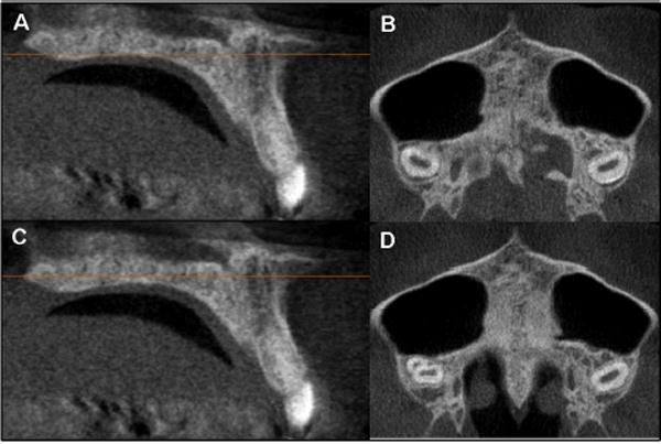
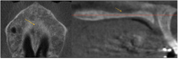
Methods: Cone-beam computed tomography images from 140 subjects (ages, 5.6-58.4 years) were examined to define the radiographic stages of midpalatal suture maturation. Five stages of maturation of the midpalatal suture were identified and defined: stage A, straight high-density sutural line, with no or little interdigitation; stage B, scalloped appearance of the high-density sutural line; stage C, 2 parallel, scalloped, high-density lines that were close to each other, separated in some areas by small low-density spaces; stage D, fusion completed in the palatine bone, with no evidence of a suture; and stage E, fusion anteriorly in the maxilla. Intraexaminer and interexaminer agreements were evaluated by weighted kappa tests.
Results: Stages A and B typically were observed up to 13 years of age, whereas stage C was noted primarily from 11 to 17 years but occasionally in younger and older age groups. Fusion of the palatine (stage D) and maxillary (stage E) regions of the midpalatal suture was completed after 11 years only in girls. From 14 to 17 years, 3 of 13 (23%) boys showed fusion only in the palatine bone (stage D).
Conclusions: This new classification method has the potential to avoid the side effects of rapid maxillary expansion failure or unnecessary surgically assisted rapid maxillary expansion for late adolescents and young adults.
Copyright © 2013 American Association of Orthodontists. Published by Mosby, Inc. All rights reserved.
Conflict of interest statement
All authors have completed and submitted the ICMJE Form for Disclosure of Potential Conflicts of Interest, and none were reported.
Figures
References
-
- Bishara SE, Staley RN. Maxillary expansion: clinical implications. Am J Orthod Dentofacial Orthop. 1987;91:3–14. - PubMed
-
- Guest SS, McNamara JA, Jr, Baccetti T, Franchi L. Improving Class II malocclusion as a side-effect of rapid maxillary expansion: a prospective clinical study. Am J Orthod Dentofacial Orthop. 2010;138:582–91. - PubMed
-
- Da Silva Filho OG, Magro AC, Capelozza Filho L. Early treatment of the Class III malocclusion with rapid maxillary expansion and maxillary protraction. Am J Orthod Dentofacial Orthop. 1998;113:196–203. - PubMed
-
- McNamara JA, Jr, Brudon WL. Orthodontics and dentofacial orthopedics. Ann Arbor Mich: Needham Press; 2001.
-
- McNamara JA., Jr Long-term adaptation to changes in the transverse dimension in children and adolescents: an overview. Am J Orthod Dentofacial Orthop. 2006;129(Suppl):S71–4. - PubMed
Publication types
MeSH terms
Grants and funding
LinkOut - more resources
Full Text Sources
Other Literature Sources
Medical
