Review
. 2013 Oct 30;80(3):568-77.
doi: 10.1016/j.neuron.2013.10.032.

Engineering approaches to illuminating brain structure and dynamics

Affiliations
Review

Engineering approaches to illuminating brain structure and dynamics

Karl Deisseroth et al. Neuron. .

Abstract

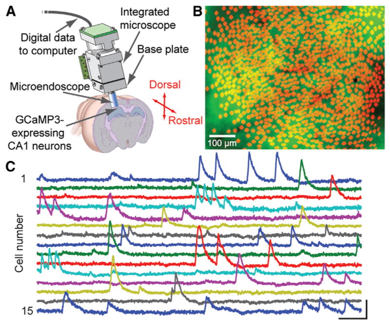
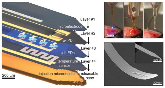
Historical milestones in neuroscience have come in diverse forms, ranging from the resolution of specific biological mysteries via creative experimentation to broad technological advances allowing neuroscientists to ask new kinds of questions. The continuous development of tools is driven with a special necessity by the complexity, fragility, and inaccessibility of intact nervous systems, such that inventive technique development and application drawing upon engineering and the applied sciences has long been essential to neuroscience. Here we highlight recent technological directions in neuroscience spurred by progress in optical, electrical, mechanical, chemical, and biological engineering. These research areas are poised for rapid growth and will likely be central to the practice of neuroscience well into the future.


Figures

Figure 1
Figure 1. Optics and Protein Engineering Converge for Ca2+ Imaging in >1,200 CA1 Pyramidal Cells in Freely Moving Mice
(A) An integrated microscope is equipped with a microendoscope to image CA1 neurons expressing the engineered Ca2+ indicator GCaMP3 under control of the Camk2a promoter. The base plate and microendoscope are fixed to the cranium for repeated access to the same field of view. Republished from Ziv et al., 2013. (B) Shown are 1,202 CA1 pyramidal cells (red somata) identified by Ca2+ imaging in a freely moving mouse atop a mean fluorescence image (green) of CA1. Vessels appear as dark shadows. Image courtesy of Yaniv Ziv and Lacey Kitch, Stanford University. (C) Example traces of [Ca2+]i dynamics from 15 cells. Scale bars denote 5% ΔF/F (vertical) and 10 s (horizontal).
Figure 2
Figure 2. Materials Science and Optoelectronics Converge for Neural Interface Research
Left: exploded-view layout of injectable semiconductor device for integrated stimulation/sensing/actuation, highlighting distinct layers for electrophysiological measurement (1), optical measurement (2), optical stimulation (3, micro-ILED array), and temperature sensing (4), all bonded to a releasable base for injection with a microneedle. Top right: injection and release of the microneedle. After insertion (left), artificial cerebrospinal fluid dissolves an external silk-based adhesive (middle) and the microneedle can be removed (right), leaving the active device in the brain. Bottom right: SEM of an injectable micro-LED array, 8.5 μm thick; flexible and rigid forms shown. Adapted with permission from Kim et al., 2013.
Figure 3
Figure 3. Chemical Engineering, Bioengineering, and Photonics Give Rise to Circuit-Probing Hardware and Wetware
Top: construction of a hydrogel from within tissue (CLARITY) creates a transparent mammalian brain for intact-system anatomical analysis; adapted from Chung et al., 2013. Bottom left: 2.3Å crystal structure of the channel-rhodopsin optogenetic control tool enables directed protein engineering for enhanced interventional functionality; adapted from Kato et al., 2012. Bottom right: optogenetic neural interfaces deliver light from laser diodes or advanced LEDs; flexible fiberoptic control in freely moving mouse shown. Photo credit Inbal Goshen and Karl Deisseroth.

References

    1. Ahrens MB, Orger MB, Robson DN, Li JM, Keller PJ. Whole-brain functional imaging at cellular resolution using light-sheet microscopy. Nat Methods. 2013;10:413–420. - PubMed
    1. Akerboom J, Carreras Calderón N, Tian L, Wabnig S, Prigge M, Tolö J, Gordus A, Orger MB, Severi KE, Macklin JJ, et al. Genetically encoded calcium indicators for multi-color neural activity imaging and combination with optogenetics. Front Mol Neurosci. 2013;6:2. - PMC - PubMed
    1. Albrecht DR, Bargmann CI. High-content behavioral analysis of Caenorhabditis elegans in precise spatiotemporal chemical environments. Nat Methods. 2011;8:599–605. - PMC - PubMed
    1. Alivisatos AP, Andrews AM, Boyden ES, Chun M, Church GM, Deisseroth K, Donoghue JP, Fraser SE, Lippincott-Schwartz J, Looger LL, et al. Nanotools for neuroscience and brain activity mapping. ACS Nano. 2013;7:1850–1866. - PMC - PubMed
    1. Anikeeva P, Andalman AS, Witten I, Warden M, Goshen I, Grosenick L, Gunaydin LA, Frank LM, Deisseroth K. Optetrode: a multi-channel readout for optogenetic control in freely moving mice. Nat Neurosci. 2012;15:163–170. - PMC - PubMed

Publication types