Review
. 2013 Oct 30;80(3):633-47.
doi: 10.1016/j.neuron.2013.10.045.

Cortical evolution: judge the brain by its cover

Affiliations
Review

Cortical evolution: judge the brain by its cover

Daniel H Geschwind et al. Neuron. .

Abstract

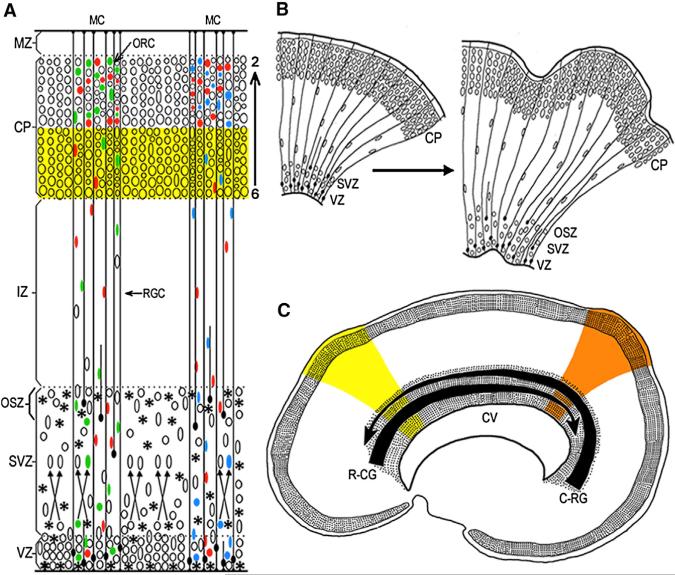
To understand the emergence of human higher cognition, we must understand its biological substrate--the cerebral cortex, which considers itself the crowning achievement of evolution. Here, we describe how advances in developmental neurobiology, coupled with those in genetics, including adaptive protein evolution via gene duplications and the emergence of novel regulatory elements, can provide insights into the evolutionary mechanisms culminating in the human cerebrum. Given that the massive expansion of the cortical surface and elaboration of its connections in humans originates from developmental events, understanding the genetic regulation of cell number, neuronal migration to proper layers, columns, and regions, and ultimately their differentiation into specific phenotypes, is critical. The pre- and postnatal environment also interacts with the cellular substrate to yield a basic network that is refined via selection and elimination of synaptic connections, a process that is prolonged in humans. This knowledge provides essential insight into the pathogenesis of human-specific neuropsychiatric disorders.


Figures

Figure 1
Figure 1. Radial Unit Model of the Deployment of Postmitotic Migratory Neurons and Their Settling Pattern into the Horizontal-Laminar, Inside-Out, and Vertical-Columnar Organization
(A) Neuronal progenitors in the proliferative ventricular and subventricular zones (VZ/SVZ/OSZ) and their progenies exhibit clonal heterogeneity (indicated by the differently colored ellipses). Several clones become intermixed in the SVZ, before migrating across the intermediate zone (IZ) along elongated shafts of the radial glial cells (RGC) into the cortical plate (CP). Newborn neurons bypass previously generated cells of the deeper layers (yellow stripe) in the inside-out sequence (layers 6 to 2) to participate in pheno-typically and functionally heterogeneous mini-columns (MC) consisting of several ontogenetic radial columns (ORC) (e.g., Rakic, 1988; Torii et al., 2009). (B) Graphic explanation of the Radial Unit Hypothesis of cortical expansion, by either preventing programmed cell death or increasing the rate of proliferation in mice, can produce a larger number of radial units that, constrained by the radial glial scaffolding, generate an expanded cellular sheet, which begins to buckle and transforms a lissencephalic (on the left) to the gyrencephalic (on the right) cerebrum. Based on studies in primates and experiments in mice (e.g., Kuida et al., 1998; Rakic, 2009; Haydar et al., 2003; Chenn and Walsh, 2003). (C) Illustration of the concept, how opposing rostro-caudal (R-CG) and caudo-rostral (C-RG) molecular gradients, that form the protomap in embryonic VZ/SVZ lining cerebral ventricle (CV) can introduce new subtypes of neurons that migrate to the overlying CP and establish new cortical areas in the superjacent CP, indicated by yellow and orange color stripes. Based on experimental data in mice (e.g., Grove and Fukuchi-Shimogori, 2003; O'Leary and Borngasser, 2006; Cholfin and Rubenstein, 2008).
Figure 2
Figure 2. Gene Coexpression Modules and Hub Genes in the Developing Human and Macaque Monkey Cerebral Cortex
(A and B) The average scaled expression of all genes in two coexpression modules with gradient-like patterns. (A) Genes in M91 exhibit a pattern with graded expression along the anterior-posterior axis. (B) Genes in M13 show a gradient in the neocortical areas of the temporal lobe. (C and D) Radar charts with qRT-PCR data of hub genes in modules 91 and 13. (C) Areal expression of CLMP (hub gene of M91) demonstrates a clear gradient-like expression pattern in humans (blue), with a graded expression from the frontal lobe to the occipital lobe. This gradient-like expression is not present in the macaque monkey (red). (D) Areal expression of NR2F2 (hub gene of M13) exhibits a gradient-like expression pattern that is conserved between the human (blue) and macaque monkey (red) temporal cortices. Based on Pletikos et al. (2013).

References

    1. Abrahams B, Tentler D, Perederiy J, Oldham M, Coppola G, Geschwind D. Genome-wide analyses of human perisylvian cerebral cortical patterning. Proc. Natl. Acad. Sci. USA. 2007;104:17849–17854. - PMC - PubMed
    1. Amato S, Liu X, Zheng B, Cantley L, Rakic P, Man HY. AMP-activated protein kinase regulates neuronal polarization by interfering with PI 3-kinase localization. Science. 2011;332:247–251. - PMC - PubMed
    1. Anderson SA, Marín O, Horn C, Jennings K, Rubenstein JL. Distinct cortical migrations from the medial and lateral ganglionic eminences. Development. 2001;128:353–363. - PubMed
    1. Anton ES, Kreidberg J, Rakic P. Distinct functions of alpha3 and alpha(v) integrin receptors in neuronal migration and laminarorganization of the cerebral cortex. Neuron. 1999;22:227–289. - PubMed
    1. Arimatsu Y, Miyamoto M, Nihonmatsu I, Hirata K, Uratani Y, Hatanaka Y, Takiguchi-Hayashi K. Early regional specification for a molecular neuronal phenotype in the rat neocortex. Proc. Natl. Acad. Sci. USA. 1992;89:8879–8883. - PMC - PubMed

Publication types