Review
. 2013 Oct 30;80(3):718-28.
doi: 10.1016/j.neuron.2013.09.044.

Homeostatic signaling and the stabilization of neural function

Affiliations
Review

Homeostatic signaling and the stabilization of neural function

Graeme W Davis. Neuron. .

Abstract

The brain is astonishing in its complexity and capacity for change. This has fascinated scientists for more than a century, filling the pages of this journal for the past 25 years. But a paradigm shift is underway. It seems likely that the plasticity that drives our ability to learn and remember can only be meaningful in the context of otherwise stable, reproducible, and predictable baseline neural function. Without the existence of potent mechanisms that stabilize neural function, our capacity to learn and remember would be lost in the chaos of daily experiential change. This underscores two great mysteries in neuroscience. How are the functional properties of individual neurons and neural circuits stably maintained throughout life? And, in the face of potent stabilizing mechanisms, how can neural circuitry be modified during neural development, learning, and memory? Answers are emerging in the rapidly developing field of homeostatic plasticity.


Figures

Figure 1
Figure 1. Evidence for the homeostatic control of cellular excitation
Top) The firing properties of central neurons are determined by a balance of synaptic excitation (red vesicles and red receptors), synaptic inhibition (blue vesicles and blue receptors), and the densities of ion channels that mediate either cellular depolarization (red ovals) or that oppose cellular depolarization (blue ovals). In response to chronic suppression of neural activity, central neurons can alter the relative abundance of ion channels and receptors at the cell surface to reestablish a set point level of activity. Bottom) At the neuromuscular junction (NMJ), chronic impairment of postsynaptic neurotransmitter receptor sensitivity or receptor abundance leads to a compensatory increase in presynaptic neurotransmitter release that precisely counteracts the change in receptor function to achieve normal synaptic depolarization of the muscle. Modified from Davis, 2006.
Figure 2
Figure 2. Accurate restoration of cellular activity through homeostatic signaling
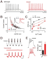
A) Sample traces from cortical pyramidal neurons from wild type and Kv4.2 knockout mice (Nerbonne et al., 2008, data from figure 8 therein). The knockout mice lack the Kv4.2 protein and current. Although acute pharmacological inhibition severely potentiates neuronal excitability, homeostatic rebalancing of potassium channel expression accurately restores firing properties to wild type levels. B) Data are shown for recordings made at the Drosophila NMJ. Presynaptic release (quantal content) is plotted against spontaneous miniature amplitudes (mEPSP). Each data point is average data from a single NMJ from control NMJ (open black) or NMJ to which philanthotoxin 433 (PhTX) was applied for 10min prior to recording (open red). The line represents ideal homeostatic compensation where any change in mEPSP is offset by an identical percent change in quantal content. The modulation of presynaptic release accurately offsets a broad range of postsynaptic perturbation. C) Data are presented for the Drosophila NMJ plotting excitatory postsynaptic current (EPSC) amplitude versus extracellular calcium concentration. Larvae treated with PhTx (wt + PhTX) accurately retarget control (wt) EPSC amplitudes across an order of magnitude change in extracellular calcium. Animals harboring a loss of function mutation in rim show reduced EPSCs at all calcium concentrations. Application of PhTX to rim mutant larvae demonstrates a failure of homeostatic compensation at all calcium concentrations (Muller et al., 2012). D) Intracellular recordings from a stomatogastric neuron in the intact ganglion (control), following removal of the ganglion and placement in organ culture for 10 minutes (Decentralized) and after four days in culture (4 days). After 4 days, the firing properties of the identified neuron are remarkably similar to that observed in the intact animal. Scale bars 1 sec / 10mV. Data modified from Khorkova and Golowasch, 2007. E) Example traces from Xenopus central neurons including control and a neuron expressing transgenic Kir2.1 (Pratt et al., 2007). Expression of Kir2.1 induces a change in the underlying current densities including the sodium current (quantified at right) that help sustain firing properties at control levels.
Figure 3
Figure 3. Cell autonomous and synapse specific homeostatic plasticity
A) Experimental configuration is diagrammed for stimulation and simultaneous recording from adjacent CA1 pyramidal neurons in hippocampal organotypic culture that are either untransfected (Record:Ctrl, black) or transfected with channel rhodopsin 2 and photostimulated in slice culture for 24hours, 50ms light pulses at 3Hz (Record: Photostim, blue). Below, representative data are shown for AMPA-mediated currents. Below is a scatter plot of recording pairs with the mean shown in red. Photostimulation causes a decrease in AMPA current due to downscaling and a decrease in synapse number (Goold and Nicoll, 2010). B) Two neighboring spines with or without overlay of the Syn-YFP terminals from a Syn-YFP/Kir2.1- overexpressing cell. 2P uncaging of MNI-glutamate was elicited at the tip of these spines (yellow crossed lines) and the resulting AMPAR-mediated synaptic current (2P-EPSC) is shown (Vh = −60 mV) (Béïque et al., 2011). A postsynaptic potentiation is seen in response to synapse specific presynaptic expression of Kir2.1. Data and images take from Goold and Nicoll (2010, A) and Béïque et al., (2011, B).
Figure 4
Figure 4. Presynaptic homeostasis is achieved by parallel changes in the size of the readily releasable vesicle pool and presynaptic calcium influx
A–B) Variance-mean analysis supports a RIM-dependent modulation of the RRP during synaptic homeostasis. Left, example EPSC traces for a WT NMJ at the indicated extracellular calcium concentration (millimolar). B) Example EPSC amplitude variance-mean plots of two WT synapses in the absence (gray) and presence (black) of PhTX with parabola fits (solid lines) that were extrapolated to the x-intercept (dashed lines; see Muller et al., 2012). C) Representative traces for measurement of the spatially averaged calcium signal within synaptic boutons at the NMJ in a wild type (control) and GluRIIA mutant. The homeostatic enhancement of presynaptic release is correlated with a statistically significant increase in the peak amplitude of the spatially averaged calcium signal (see Muller and Davis, 2011).
Figure 5
Figure 5. Emerging model for the expression of presynaptic homeostasis
Schematic of an active zone at the Drosophila NMJ shown presynaptic CaV2.1 calcium channels (blue), the action potential triggered calcium microdomain (red), postsynaptic glutamate receptors (GluRIIA, dark gray) and presynaptic ENaC channel (pink). Two genes, pickpocket11 and pickpocket16 were discovered to be necessary, presynaptically, for the rapid induction and sustained expression of presynaptic homeostasis (Younger et al., 2013). These genes encode subunits of an Epithelial/Degenerin (ENaC) sodium leak channel. These genes are co-transcribed and transcriptionally upregulated following persistent disruption of postsynaptic glutamate receptor function. These and other data support a model in which ENaC channel insertion drives a modest depolarization of the presynaptic resting potential (ΔV), which enhances presynaptic calcium and neurotransmitter release. A parallel change in the readily releasable pool of vesicles is also necessary for synaptic homeostasis, which relies on the presynaptic adaptor proteins RIM (Muller et al., 2012) and RIM binding protein (RBP) (green). When both process are enabled, a homeostatic enhancement of release is observed. A number of additional synaptic proteins have been shown to be required for presynaptic homeostasis (dark blue text). Among them, there is evidence for the involvement of micro-RNA-based signaling (Mir10, gray; Tsurudome et al., 2011) and permissive BMP mediated signaling released from muscle (gray) and acting at the motoneuron soma (gray, Goold et al., 2007). Postsynaptically, there is evidence for the required function of Tor and S6K as well as Dystrobrevin-dependent scaffolding (Penney et al,. 2012; Pilgram et al., 2011). Figure modified from Younger et al., (2013). Additional molecular mechanisms involved in presynaptic homeostasis have been recently reviewed (Frank, 2013).

Similar articles

Cited by

References

    1. Andrásfalvy BK, Makara JK, Johnston D, Magee JC. Altered synaptic and non-synaptic properties of CA1 pyramidal neurons in Kv4.2 knockout mice. J Neurophysiol. 2008;586:3881–3892. - PMC - PubMed
    1. Anggono V, Clem RL, Huganir RL. PICK1 loss of function occludes homeostatic synaptic scaling. J Neurosci. 2011;31:2188–2196. - PMC - PubMed
    1. Awatramani GB, Price GD, Trussell LO. Modulation of Transmitter Release by Presynaptic Resting Potential and Background Calcium Levels. Neuron. 2005;48:109–121. - PubMed
    1. Bateup HS, Johnson CA, Denefrio CL, Saulnier JL, Kornacker K, Sabatini BL. Excitatory/inhibitory synaptic imbalance leads to hippocampal hyperexcitability in mouse models of tuberous sclerosis. Neuron. 2013;78:510–522. - PMC - PubMed
    1. Baumgardt M, Miguel-Aliaga I, Karlsson D, Ekman H, Thor S. Specification of neuronal identities by feedforward combinatorial coding. PLoS biology. 2007;5:e37. - PMC - PubMed

Publication types