Review
. 2014 Feb;163(2):109-22.
doi: 10.1016/j.trsl.2013.10.003. Epub 2013 Oct 14.

New tides: using zebrafish to study renal regeneration

Affiliations
Review

New tides: using zebrafish to study renal regeneration

Kristen K McCampbell et al. Transl Res. 2014 Feb.

Abstract

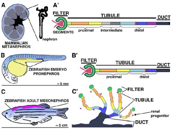
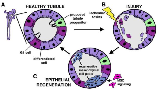
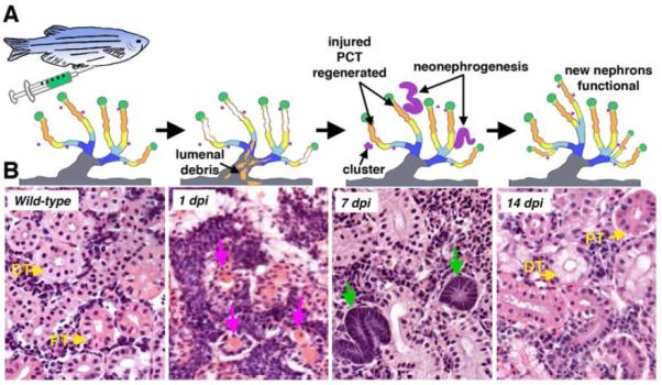
Over the past several decades, the zebrafish has become one of the major vertebrate model organisms used in biomedical research. In this arena, the zebrafish has emerged as an applicable system for the study of kidney diseases and renal regeneration. The relevance of the zebrafish model for nephrology research has been increasingly appreciated as the understanding of zebrafish kidney structure, ontogeny, and the response to damage has steadily expanded. Recent studies have documented the amazing regenerative characteristics of the zebrafish kidney, which include the ability to replace epithelial populations after acute injury and to grow new renal functional units, termed nephrons. Here we discuss how nephron composition is conserved between zebrafish and mammals, and highlight how recent findings from zebrafish studies utilizing transgenic technologies and chemical genetics can complement traditional murine approaches in the effort to dissect how the kidney responds to acute damage and identify therapeutics that enhance human renal regeneration.


Figures

Fig 1
Fig 1. Kidney architecture varies between vertebrates, but the strategy for nephron segmental composition is broadly conserved
(A) The prototypical mammalian mesonephros is a kidney composed of nephrons ranging in number from thousands to millions, and each nephron is an epithelial tubes with a macroscopic structure of stereotypical loops and convolutions. (A’) The nephron is drawn as a straightened epithelial tube. It is comprised of a filter, tubule and duct, with a regular pattern of proximal, intermediate, and distal segments of epithelial cells that have discrete roles in modifying the filtrate during urine production. The mammalian nephron segments are as follows: blood filter (dark green surrounding red capillary network); neck (light green); proximal convoluted tubule (orange); proximal straight tubule (yellow); descending thin limb (light grey); ascending thin limb (dark grey); thick ascending limb (light blue); macula densa (red); distal convoluted tubule (dark blue); connecting tubule (purple); collecting duct (black). (B) The zebrafish embryo initially develops a linear pronephros, with a pair of nephrons, and laterally a single nephron can be visualized. (B’) The zebrafish pronephric nephron has a blood filter, multi-segmented tubule, and duct. Analogous segments to the mammalian nephron are indicated by color. The pronephros nephron segments are as follows: blood filter (dark green surrounding red capillary network); neck (light green); proximal convoluted tubule (orange); proximal straight tubule (yellow); distal early (light blue); Corpuscle of Stannius (red); distal late (dark blue); collecting duct (black). (C) The zebrafish adult contains a single, flattened mesonephric kidney on the dorsal wall of the body cavity. (C’) Examination of the mesonephros nephron arrangement and constitution has found that nephrons are arranged in branched units and pinwheel-like arrays that connect to a central duct system. The mesonephric nephrons have similar segments as the embryonic nephrons, and intervening stroma contains renal progenitors that can form new nephrons after injury. The mesonephros nephron segments are as follows: blood filter (dark green circle); neck (light green); proximal convoluted tubule (orange); proximal straight tubule (yellow); distal early (light blue); distal late (dark blue); collecting duct (black).
Fig 2
Fig 2. Comparison of renal histology between zebrafish and mouse
(A) Zebrafish mesonephros and (B) mouse metanephros sections stained with hematoxylin and eosin. (A) This zebrafish section includes proximal tubule (PT) (dark pink) and distal tubule (DT) (light pink) cross-sections along with a dense interstitial stroma (arrowheads) with intensely-purple stained nuclei that includes hematopoietic cells and is also the proposed location of renal progenitors. This particular (B) mouse section is dominated by distal tubules (DT) (light pink).
Fig 3
Fig 3. Acute kidney injury
(A) (Left) A schematic depiction of a nephron, with the level of cross section indicated. (Right) Healthy tubule cross-section, with differentiated epithelial cells (purple) interspersed with currently debated regeneration sources: cells in G1 undergoing low rate of turnover (magenta), and the renal progenitor/stem cell (light green). (B) Schematic after injury, with luminal debris and surviving epithelial cells. (C) During renal epithelial regeneration, mesenchymal cells (blue) have been observed within the tubule. Whether these mesenchymal cells emerge from renal progenitors/stem cells that reside in the nephron, from differentiated cells (either in G1 or non-cycling) that dedifferentiate, or some combination of these sources, is currently an active area of nephrology research. Cells located in the interstitial space between nephrons that can impact tubular regeneration include MSCs (pink). After the tubular regenerative cells proliferate, their offspring differentiate and re-establish tubular integrity.
Fig 4
Fig 4. AKI modeling in the zebrafish embryo
Gentamicin exposure resulted in pericardial edema and hindered proximal tubule development. (A) Live images of a wild-type and embryos injected with gentamicin at 48 hpf. The wild-type embryo and 24 hpi embryo shown were 72 hpf, with 48 and 72 hpi embryos shown to document the progression of the gentamicin phenotype. Gentamicin injected embryos displayed a darkened yolk sac and pericardial edema at 24-72 hpi. 4X magnification. (B) A comparison of whole mount in situ hybridization of embryos following either dextran injection (vehicle control) or gentamicin injection at 48 hpf. Gentamicin injected embryos showed delayed PCT (arrow) coiling, visualized with the slc20a1a transcript signal (purple); asterisk (*) indicates slc20a1a staining in trunk mesenchyme that is not associated with the pronephros. Blue arrowheads demarcate tubular folds on the left nephron in 48 and 72 hpi embryos, the number of which is reduced in gentamicin-injected embryos. Dorsal views are shown, with anterior to the left. 10X magnification.
Fig 5
Fig 5. Analysis of tubule cell composition and architecture revealed that gentamicin disrupts the apical-basal polarity of renal tubules
Tubules of wild-type enpep:eGFP transgenic embryos and gentamicin injected embryos were analyzed with immunohistochemistry to detect the tubule cells, which were demarcated using anti-eGFP antibody (green). The apical surface of tubule cells was labeled with phalloidin (red) and nuclei were labeled with DAPI (blue). Control embryos at 3 dpf and 4 dpf displayed an intact luminal border, whereas embryos that received gentamicin showed structural disruptions in the phalloidin staining of tubular epithelial cells at 24 hpi (white arrowhead), and displayed collapsed lumens at 48 hpi (white arrows). 60X magnification. All embryos were injected at 48 hpf.
Fig 6
Fig 6. Laser ablation of the zebrafish pronephros
(A-) (Top) zebrafish schematic showing region of proximal tubule laser ablation, with (bottom) tubule labeled with dextran-FITC (green) or dextran-rhodamine (red). Cells ablated at day 3 of development are replaced by day 7, suggestive of robust tubular proliferation that regenerates the ablated cell populace. (B) Whole mount in situ hybridization for slc20a1a to demarcate the proximal tubule in a wild-type control (left), or embryos fixed immediately after ablation: an embryo with extensive cell ablation (center) and an embryo with a focal ablation (right). All views are dorsal. *Images reprinted with permission.
Fig 7
Fig 7. AKI modeling in the zebrafish adult: there are two regeneration responses
(A) Intraperitoneal injection of gentamicin into the adult fish (schematic). (B) (Top) A timecourse of schematics and (bottom) histological sections stained with hematoxylin and eosin showing the major cellular events. The uninjured kidney contains both proximal tubules (PT) and distal tubules (DT) (yellow arrows). At 1 day post injection (dpi), luminal debris is seen as tubular casts (pink arrows) that fill surviving tubule lumens. At 7 dpi, proximal convoluted tubule (PCT) integrity is restored, and sections contain basophilic (dark purple) clusters and S-shaped tubular structures that correspond to new nephrons (green arrows). By 14 dpi, basophilic structures are infrequent, and the tissue is dominated by tubules with either proximal (dark pink) or distal (light pink) staining (yellow arrows).

Similar articles

Cited by

References

    1. Reilly RF, Bulger RE, Kriz W. Diseases of the Kidney and Urinary Tract. Lippincott Williams & Wilkins; Philadelphia: 2007. Structural-functional relationships in the kidney; pp. 2–53.
    1. Guyton AC, Hall JE. Textbook of Medical Physiology. Elsevier Saunders; Philadelphia: 2006. p. 310.
    1. Dressler GR. The cellular basis of kidney development. Annu Rev Cell Dev Biol. 2006;22:509–529. - PubMed
    1. Vize PD, Seufert DW, Carroll TJ, Wallingford JB. Model systems for the study of kidney development: use of the pronephros in the analysis of organ induction and patterning. Dev Biol. 1997;188:189–204. - PubMed
    1. Drummond I. Making a zebrafish kidney: a tale of two tubes. Trends Cell Biol. 2003;13:357–365. - PubMed

Publication types