. 2014 Feb 1;386(1):181-90.
doi: 10.1016/j.ydbio.2013.10.026. Epub 2013 Oct 30.

Cerebellar development in the absence of Gbx function in zebrafish

Affiliations

Cerebellar development in the absence of Gbx function in zebrafish

Chen-Ying Su et al. Dev Biol. .

Abstract

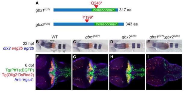
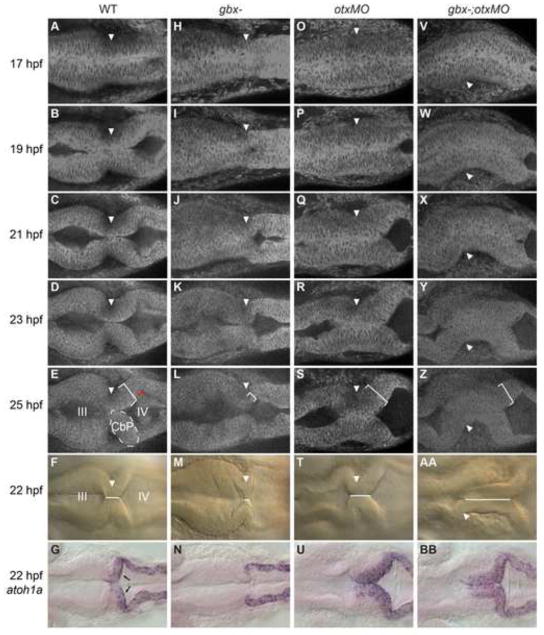
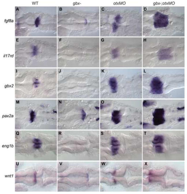
The midbrain-hindbrain boundary (MHB) is a well-known organizing center during vertebrate brain development. The MHB forms at the expression boundary of Otx2 and Gbx2, mutually repressive homeodomain transcription factors expressed in the midbrain/forebrain and anterior hindbrain, respectively. The genetic hierarchy of gene expression at the MHB is complex, involving multiple positive and negative feedback loops that result in the establishment of non-overlapping domains of Wnt1 and Fgf8 on either side of the boundary and the consequent specification of the cerebellum. The cerebellum derives from the dorsal part of the anterior-most hindbrain segment, rhombomere 1 (r1), which undergoes a distinctive morphogenesis to give rise to the cerebellar primordium within which the various cerebellar neuron types are specified. Previous studies in the mouse have shown that Gbx2 is essential for cerebellar development. Using zebrafish mutants we show here that in the zebrafish gbx1 and gbx2 are required redundantly for morphogenesis of the cerebellar primordium and subsequent cerebellar differentiation, but that this requirement is alleviated by knocking down Otx. Expression of fgf8, wnt1 and the entire MHB genetic program is progressively lost in gbx1-;gbx2- double mutants but is rescued by Otx knock-down. This rescue of the MHB genetic program depends on rescued Fgf signaling, however the rescue of cerebellar primordium morphogenesis is independent of both Gbx and Fgf. Based on our findings we propose a revised model for the role of Gbx in cerebellar development.

Keywords: Cerebellum; Gbx; Zebrafish.


Figures

Fig. 1
Fig. 1. Gbx1 and Gbx2 function redundantly in cerebellum development
(A) Schematic of nonsense mutations identified in zebrafish gbx1 and gbx2 by TILLING. Both mutations are expected to prevent DNA binding by truncating the homeodomain. (B–I) Dorsal views at 22 hpf (B–E) or 6 dpf (F–I), anterior to the left. Genotypes are shown at the top. otx2 (blue), eng2b (red), and egr2b (blue) are expressed in midbrain, midbrain-hindbrain boundary (MHB), and rhombmere 3/5, respectively, in wildtype (WT), single and double mutants as shown. Tg(ptf1a:EGFP) (green) marks Purkinje neuron progenitors, Tg(olig2:DsRed2) (red) marks projection neurons, and anti-Vglut1/Slc17a7 (blue) marks cerebellar granule neuron axons. In gbx1fh271;gbx2fh253 double mutants the midbrain is expanded at the expense of r1 and no cerebellum forms.
Fig. 2
Fig. 2. Cerebellar development is rescued in gbx- embryos with otx knock-down
Dorsal views at 5 dpf, anterior is to the left. Genotypes are indicated at the top. (A–H) Zebrin II/Aldoca (A–D) and Vglut1/Slc17a7 (E–H) are expressed in cerebellar Purkinje cells and granule cell axons respectively; both are absent in gbx- and rescued in otxMO and gbx-;otxMO.
Fig. 3
Fig. 3. Rescue of cerebellar primordium morphogenesis in gbx- embryos with otx knock-down
Timelapse of embryos during the initial stages of cerebellar morphogenesis. Dorsal views with anterior to the left; genotypes are indicated at the top. Arrowheads indicate the isthmic constriction. (A–E) In WT embryos, inflation of the midbrain (III) and hindbrain (IV) ventricles combined with cell shape changes at the MHB creates a sharp isthmic constriction flanked posteriorly by a thickened bilateral cerebellar primordium (CbP, indicated on the left side with dotted white lines and on the right side by a white bracket indicating thickness). Red lines indicate a 90° rotation of the CbP relative to the more posterior hindbrain epithelium. (H–L) Ventricle inflation is delayed in gbx mutants, the isthmic region is short and no thickened CbP forms. (O–S;V–Z) In otxMO and gbx-;otxMO the isthmic region is extended and a thickened CbP forms. (F,M,T,AA) The “isthmic region” is where the left and right sides of the neuroepithelium are in contact (white line), and can be easily measured at 22 hpf in WT (F). This region is nearly absent in gbx- (M), but extended in otxMO (T) and gbx-;otxMO (AA). (G,N,U,BB) atoh1a expression in granule cell progenitors in the upper rhombic lip (URL, black arrows) is lost in gbx- (N) and rescued in both otxMO (U) and gbx-;otxMO (BB).
Fig. 4
Fig. 4. Rescue of the MHB program in gbx- embryos with otx knock-down
RNA in situ hybridization with genes shown on left; genotypes indicated at the top. Dorsal views of 22 hpf embryos with anterior to the left. (A–T) fgf8a, il17rd/sef, gbx2, pax2a and eng1b are all expressed at or around the MHB, are absent or strongly reduced in gbx- but are expanded in both otxMO and gbx-;otxMO embryos. (U–X) wnt1 is normally expressed in a narrow domain anterior to the MHB (U). This expression is reduced in gbx- (V) but rescued anterior to the extended isthmic region in otxMO (W) and gbx-;otxMO (X).
Fig. 5
Fig. 5. Changing requirements for wnt1 and fgf8 expression leads to rescue of a wnt1-fgf8 boundary in gbx-;otxMO embryos
RNA in situ hybridizations with wnt1 (red) and fgf8a (blue) at the stages shown on the left; genotypes indicated at the top. Dorsal views with anterior to the left. (A–D,F–I,K–N) At 10.33 hpf (1–3 somites) in WT (A), wnt1 (red) and fgf8a (blue) are expressed in broad domains anterior and posterior to the presumptive MHB respectively (black arrow indicates the fgf8a domain in the anterior hindbrain). fgf8a is also expressed in hindbrain rhombomere 4 (black dot). In gbx- embryos wnt1 is initially expanded (B,G) but subsequently lost (L). Conversely, in otxMO and gbx-;otxMO, wnt1 expression is initially reduced (C,D) but subsequently recovers while the fgf8a domain expands (H,I,M,N). (E,J,O) Genetic pathways indicate the decreasing dependence of Wnt1 expression on Otx in the midbrain and its increasing dependence on Fgf8 signaling from r1.
Fig. 6
Fig. 6. Fgf signaling is required for rescue of the MHB program and cerebellar differentiation but not URL specification in gbx-;otxMO
Dorsal views of 22 hpf embryos (A–H) or 4 dpf larvae (I–L) with anterior to the left. Genotypes are indicated at the top. (A–H) RNA in situ hybridizations with genes indicated on left. atoh1a expression in the URL (black arrows in A) is absent in dnFgfr (B) but is rescued in dnFgfr;otxMO (C) and gbx-;dnFgfr;otxMO embryos (D). By contrast, MHB gene expression (pax2a, E) is not rescued by otx knock-down in the absence of Fgf signaling (G and H). (I–L) Zebrin II/Aldoca (red) normally expresses in cerebellar Purkinje cells and Tg(ptf1a:EGFP) (green) marks Purkinje neuron progenitors in WT (I; 14 larvae examined), but zebrin expression is absent and Tg(ptf1a:EGFP) is only expressed in lower rhombic lip in gbx-;dnFgfr;otxMO (L; 8 larvae examined). In dnFgfr (J) larvae, zebrin expression is absent (9 out of 18 larvae) or few zebrin-expressing cells are lying laterally at the junction of the tectum and the ptf1a:EGFP-expressing lower rhombic lip (the box in J; 9 out of 18). Similar to dnFgfr larvae, zebrin expression is either in the lateral junction between the tectum and lower rhombic lip in dnFgfr;otxMO (K; 15 out of 30 larvae) or is absent (the box in K).
Fig. 7
Fig. 7. A model for cerebellar morphogenesis
Our results suggest that normal cerebellar differentiation results from both the expression of the MHB program and the morphogenesis of the cerebellar primordium where cerebellar cell fates arise. Although our focus here has been on the URL, the CbP also includes the generative zone for Purkinje and projection neuron progenitors. Gbx and Fgf8 promote both processes by inhibiting their repression by Otx. Additionally, Fgf8 is required for the execution of the MHB program itself. Thus CbP morphogenesis and URL specification are both rescued in the absence of Gbx or Fgf8 signaling (or both) by Otx knock-down, while the MHB program and further cerebellar differentiation in Otx knock-down embryos requires Fgf8 but not Gbx.

References

    1. Acampora D, Avantaggiato V, Tuorto F, Briata P, Corte G, Simeone A. Visceral endoderm-restricted translation of Otx1 mediates recovery of Otx2 requirements for specification of anterior neural plate and normal gastrulation. Development. 1998;125:5091–104. - PubMed
    1. Bae YK, Kani S, Shimizu T, Tanabe K, Nojima H, Kimura Y, Higashijima S, Hibi M. Anatomy of zebrafish cerebellum and screen for mutations affecting its development. Dev Biol. 2009;330:406–26. - PubMed
    1. Bally-Cuif L, Cholley B, Wassef M. Involvement of Wnt-1 in the formation of the mes/metencephalic boundary. Mech Dev. 1995;53:23–34. - PubMed
    1. Broccoli V, Boncinelli E, Wurst W. The caudal limit of Otx2 expression positions the isthmic organizer. Nature. 1999;401:164–8. - PubMed
    1. Buckley DM, Burroughs-Garcia J, Lewandoski M, Waters ST. Characterization of the Gbx1−/− mouse mutant: a requirement for Gbx1 in normal locomotion and sensorimotor circuit development. PLoS One. 2013;8:e56214. - PMC - PubMed

Publication types

MeSH terms

LinkOut - more resources