. 2013 Nov 18;23(22):2255-2261.
doi: 10.1016/j.cub.2013.09.019. Epub 2013 Oct 31.

Polo-like kinase 4 autodestructs by generating its Slimb-binding phosphodegron

Affiliations

Polo-like kinase 4 autodestructs by generating its Slimb-binding phosphodegron

Joseph E Klebba et al. Curr Biol. .

Abstract

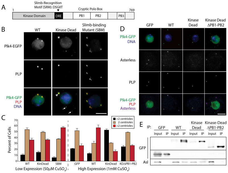
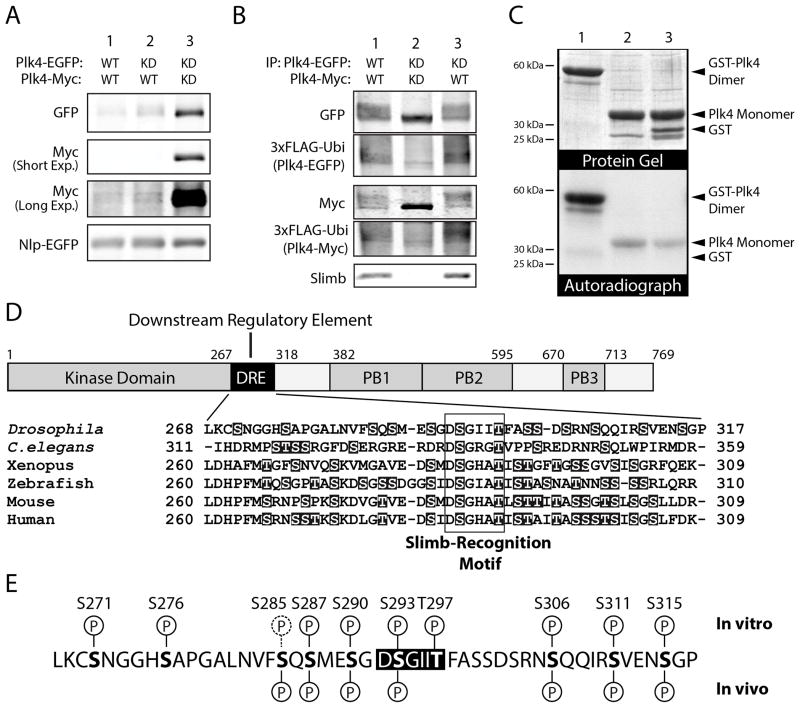
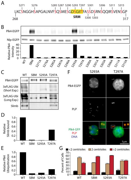
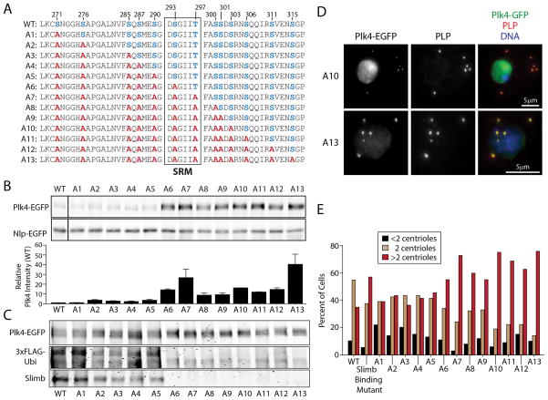
Polo-like kinase 4 (Plk4) is a conserved master regulator of centriole assembly. Previously, we found that Drosophila Plk4 protein levels are actively suppressed during interphase. Degradation of interphase Plk4 prevents centriole overduplication and is mediated by the ubiquitin-ligase complex SCF(Slimb/βTrCP). Since Plk4 stability depends on its activity, we studied the consequences of inactivating Plk4 or perturbing its phosphorylation state within its Slimb-recognition motif (SRM). Mass spectrometry of in-vitro-phosphorylated Plk4 and Plk4 purified from cells reveals that it is directly responsible for extensively autophosphorylating and generating its Slimb-binding phosphodegron. Phosphorylatable residues within this regulatory region were systematically mutated to determine their impact on Plk4 protein levels and centriole duplication when expressed in S2 cells. Notably, autophosphorylation of a single residue (Ser293) within the SRM is critical for Slimb binding and ubiquitination. Our data also demonstrate that autophosphorylation of numerous residues flanking S293 collectively contribute to establishing a high-affinity binding site for SCF(Slimb). Taken together, our findings suggest that Plk4 directly generates its own phosphodegron and can do so without the assistance of an additional kinase(s).


Figures

Figure 1
Figure 1. High Expression of Kinase-dead Plk4 Blocks Centriole Duplication by Preventing Asterless Targeting to Centrioles
(A) Linear map of Drosophila Plk4 showing functional and structural domains. The Downstream Regulatory Element (DRE) is a span of approximately 50 amino acids containing the phylogenetically conserved Slimb-recognition motif (SRM). Plk4 contains three Polo-boxes (PB) [10]. PB1 and PB2 comprise the Asl-binding region [, –22]. (B) S2 cells co-expressing the indicated Plk4-EGFP construct (green) and Nlp-EGFP (a nuclear protein used as a transfection marker; green nuclei) were immunostained for PLP (red) to mark centrioles. DNA (blue). Expression of Plk4 constructs was induced with 50μM CuSO4. KD-Plk4 targets centrioles (arrowhead) but also forms cytoplasmic punctate aggregates (arrows). Scale, 5μm. (C) Transfected S2 cells were induced to express Plk4-EGFP constructs at low (50μM CuSO4) or high (1mM CuSO4) levels for three days, immunostained for PLP, and their centrioles counted. Centriole amplification (a significant, increased frequency of >2 centrioles/cell) occurs in cells expressing SBM-Plk4 (P=0.0001) or a high level of WT-Plk4 (P=0.0002). In contrast, centriole duplication is inhibited (a significant, increased frequency of <2 centrioles/cell) in cells expressing a high level of KD-Plk4 (P<0.0001) but not KD-Plk4-ΔPB1-PB2 (P=0.29). At least three experiments were performed per construct (n = 600 cells/construct). Error bars in all figures, S.E.M. (D) S2 cells co-expressing the indicated Plk4-EGFP construct (green) and the transfection marker, Nlp-EGFP (green nuclei), were immunostained for PLP (red) and Asterless (blue; bottom row). DNA (blue; top row). Expression of Plk4 constructs was induced with 1mM CuSO4. Scale, 5μm. (E) Anti-GFP immunoprecipitates from S2 cell lysates transiently-expressing the indicated Plk4-EGFP construct (or control GFP) were probed for GFP and endogenous Asterless. Note that co-precipitating Asl is absent in the control and KDΔPB1-PB2 samples.
Figure 2
Figure 2. Plk4 is Destabilized by Trans-Autophosphorylation and Directly Autophosphorylates its SRM In Vitro
(A) The relative protein stabilities of different combinations of Plk4 constructs were analyzed by immunoblotting lysates of S2 cells transiently co-expressing the indicated EGFP- and Myc-tagged Plk4 constructs and Nlp-EGFP (used as a loading control and expressed under its endogenous promoter). Anti-Myc immunoblots are shown at short and long exposures. (B) Anti-GFP immunoprecipitates (IPs) were prepared from lysates of S2 cells transiently expressing 3xFLAG-ubiquitin and the indicated combinations of EGFP- and Myc-tagged Plk4 constructs. Blots of the IPs were probed with anti-GFP, FLAG, and Slimb antibodies. Note that robust ubiquitination of Plk4 corresponds to the presence of endogenous Slimb, and that co-expression of KD-Plk4 (lane 2) prevents phosphorylation (as indicated by the lack of gel-shift), Slimb binding, and ubiquitination. (C) Autophosphorylation of Plk4 kinase domain is more efficient as a dimer compared to a monomeric species. Lanes 1–3, both purified recombinant GST-tagged (dimeric) human Plk4 kinase domain + SRM and monomeric kinase (cleaved of its GST-tag) autophosphorylates in vitro. Plk4 does not phosphorylate purified GST (lane 3). Top panel, Coomassie-stained SDS-PAGE gel; bottom panel, corresponding autoradiograph. Equimolar amounts of dimeric and monomeric kinase were assayed. (D) The DRE contains a conserved, high percentage (~20–40%) of hydroxyl amino acids (highlighted) that are potential sites of phosphorylation. These include the conserved Ser and Thr residues within the SRM (boxed). (E) In vitro phosphorylation sites within the DRE were identified by tandem mass spectrometry (MS) analysis of purified fly His6-Plk4 (amino acid residues 1–317, comprising the kinase and DRE domains) incubated with MgATP. Above the DRE sequence, in vitro phosphorylation sites identified with high confidence are indicated with a ‘P’ encircled with a solid line; a low confidence site (S285) is indicated with a ‘P’ encircled with a dashed line. In vivo phosphorylation sites within the DRE were identified by MS analysis of full-length Plk4-EGFP immunoprecipitated from S2 cell lysates. Identified in vivo sites are marked below the DRE sequence. (A very low confidence site, S271, is not marked.) At least nine of the hydroxyl residues within the DRE are phosphorylated in vitro, and seven of these residues are also phosphorylated in vivo (bottom row).
Figure 3
Figure 3. S293 of the SRM is the Critical DRE Residue for Slimb Recognition
(A) 13 hydroxyl amino acids (red) within the DRE of full-length fly Plk4 were individually mutated to alanines and used to evaluate the impact of each residue on Plk4 stability and centriole duplication. S293 and T297 reside within the SRM (yellow highlight). (B) (Top) Anti-GFP immunoblot of lysates prepared from S2 cells transiently co-expressing the indicated Plk4-EGFP construct and Nlp-EGFP (loading control). All Plk4 mutants are single alanine mutants except SBM (a double mutant of S293A/T297A). (Bottom) Plk4-EGFP intensities were measured by densitometry of the anti-GFP immunoblot and normalized with their respective Nlp-EGFP loading controls. The plotted values are the normalized Plk4 intensities relative to the WT-Plk4 treatment. (C) Anti-GFP immunoprecipitates of lysates prepared from S2 cells transiently expressing 3xFLAG-ubiquitin and the indicated Plk4-EGFP construct were probed with anti-GFP, FLAG, and Slimb antibodies. Short and long exposures of the anti-FLAG immunoblot are shown. (D) Amounts of associated endogenous Slimb were determined by densitometry of the anti-Slimb immunoblot and then normalizing the measurements with the amounts of Plk4-EGFP present in the IPs. The plotted values are relative to the WT-Plk4 treatment. (E) The relative amounts of total Plk4 FLAG-Ubi were calculated using the densitometry method described in (D). (F) S2 cells co-expressing the indicated Plk4-EGFP construct (green puncta) and Nlp-EGFP (green nuclei) were immunostained for PLP (red) to mark centrioles. DNA (blue). Insets show higher magnifications of the boxed regions. (G) The centrioles of S2 cells treated and immunostained as in (F) were counted. Centrioles are amplified in cells expressing SBM (P<0.0001) or S293A (P<0.0001) but not T297A (P=0.06). There is no significant difference in centriole loss (black bars) in these treatments. Four experiments were performed per construct (n = 600 cells/construct).
Figure 4
Figure 4. Mutation of All 13 Hydroxyl DRE Residues Display Only a Subtle Difference in Plk4 Stability Compared to the S293A Mutant
(A) Hydroxyl residues (blue) within the DRE were systematically mutated to non-phosphorylatable alanines (red) to generate a series of Plk4 constructs containing an increasing number of mutations. (B) Plk4 is stabilized by mutation of residue S293, but this stability is slightly modulated by neighboring phosphorylatable residues within the DRE. (Top) Anti-GFP immunoblot of lysates from S2 cells transiently co-expressing the indicated Plk4-EGFP construct and Nlp-EGFP (loading control). (Bottom) Relative amounts of Plk4 protein were determined by measurement of the integrated intensities of the Plk4 bands, normalized to their respective loading controls, and then plotted relative to the normalized intensity of WT-Plk4. Measurements were obtained from three experiments. (C) Blots of anti-GFP immunoprecipitates from lysates of S2 cells transiently expressing 3xFLAG-Ubi and the indicated Plk4-EGFP construct were probed with anti-GFP, FLAG, and Slimb antibodies. Slimb binding is reduced by mutation of upstream DRE serines (A2–A5). As expected, Slimb binding is eliminated in mutants containing the S293A mutation (A6–A13). (D) S2 cells co-expressing the indicated Plk4-EGFP plasmid (green puncta) and Nlp-EGFP (green nuclei) were immunostained for PLP to mark centrioles (red). DNA (blue). (E) Expression of stabilized Plk4 mutants increases the percent of cells with excess centrioles (>2). Centrioles in 100 cells were measured per construct.

References

    1. Brito DA, Gouveia SM, Bettencourt-Dias M. Deconstructing the centriole: structure and number control. Curr Opin Cell Biol. 2012;24:4–13. - PubMed
    1. Brownlee CW, Klebba JE, Buster DW, Rogers GC. The protein phosphatase 2A regulatory subunit Twins stabilizes Plk4 to induce centriole amplification. J Cell Biol. 2011;195:231–243.3. - PMC - PubMed
    1. Rogers GC, Rusan NM, Roberts DM, Peifer M, Rogers SL. The SCFSlimb ubiquitin ligase regulates Plk4/Sak levels to block centriole reduplication. J Cell Biol. 2009;184:225–239. - PMC - PubMed
    1. Cunha-Ferreira I, Rodrigues-Martins A, Bento I, Riparbeli M, Zhang W, Laue E, Callaini G, Glover DM, Bettencourt-Dias M. The SCF/Slimb ubiquitin ligase limits centrosome amplification through degradation of SAK/PLK4. Curr Biol. 2009;19:43–49. - PubMed
    1. Holland AJ, Lan W, Niessen S, Hoover H, Cleveland DW. Polo-like kinase 4 kinase activity limits centrosome overduplication by autoregulating its own stability. J Cell Biol. 2010;188:191–198. - PMC - PubMed

Publication types

MeSH terms

LinkOut - more resources