. 2013 Nov 18;23(22):2224-2232.
doi: 10.1016/j.cub.2013.09.029. Epub 2013 Oct 31.

Polyploidization and cell fusion contribute to wound healing in the adult Drosophila epithelium

Affiliations

Polyploidization and cell fusion contribute to wound healing in the adult Drosophila epithelium

Vicki P Losick et al. Curr Biol. .

Abstract

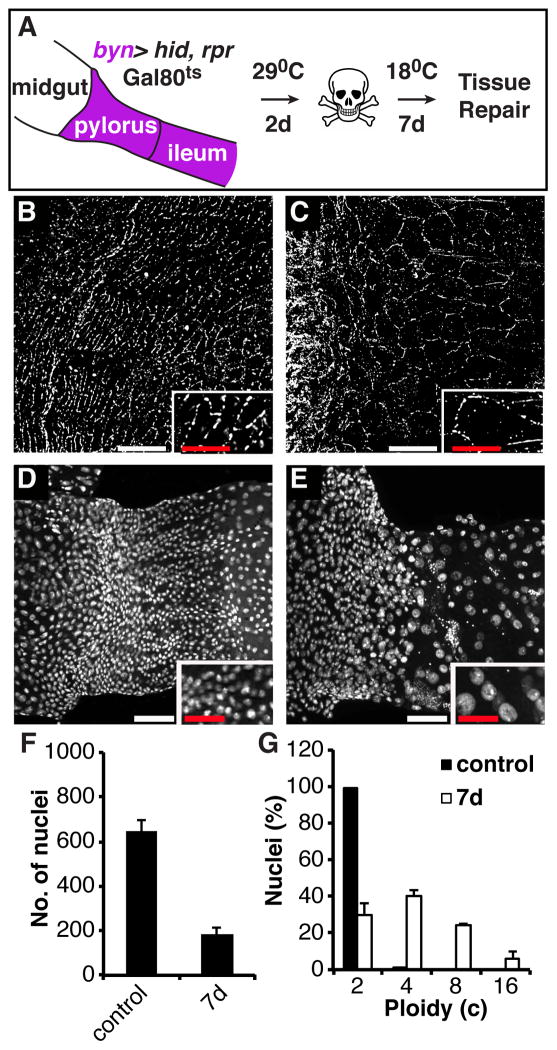
Background: Reestablishing epithelial integrity and biosynthetic capacity is critically important following tissue damage. The adult Drosophila abdominal epithelium provides an attractive new system to address how postmitotic diploid cells contribute to repair.

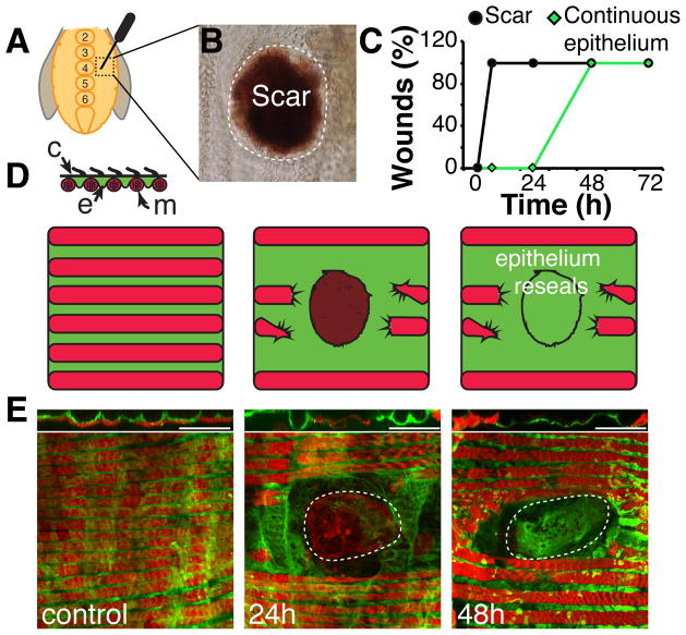
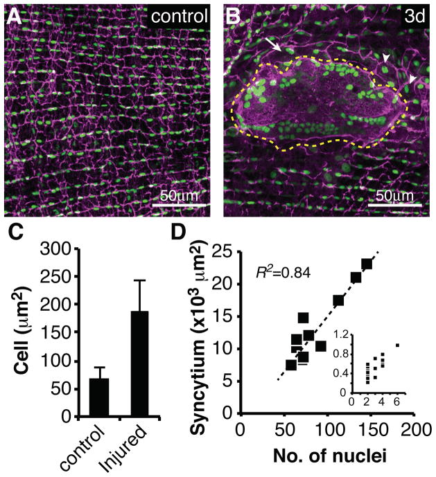
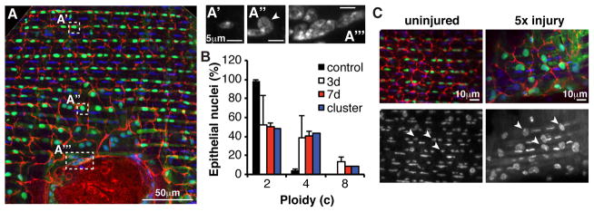
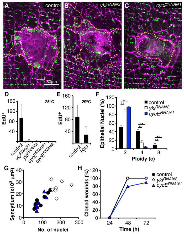
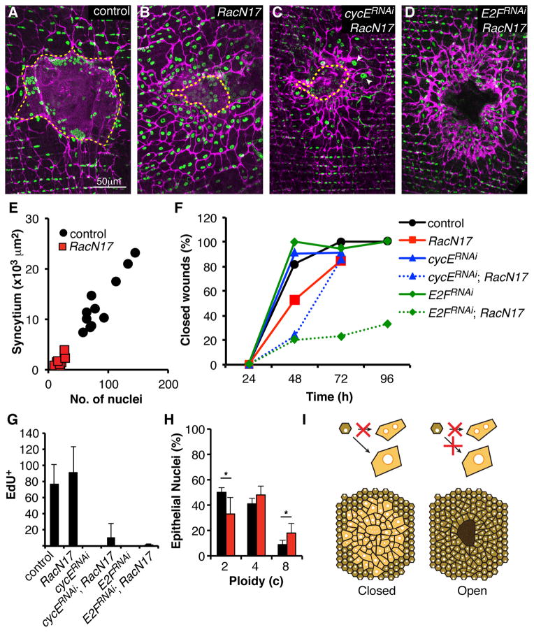
Results: Puncture wounds to the adult Drosophila epidermis close initially by forming a melanized scab. We found that epithelial cells near the wound site fuse to form a giant syncytium, which sends lamellae under the scab to re-epithelialize the damaged site. Other large cells arise more peripherally by initiating endocycles and becoming polyploid, or by cell fusion. Rac GTPase activity is needed for syncytium formation, while the Hippo signaling effector Yorkie modulates both polyploidization and cell fusion. Large cell formation is functionally important because when both polyploidization and fusion are blocked, wounds do not re-epithelialize.

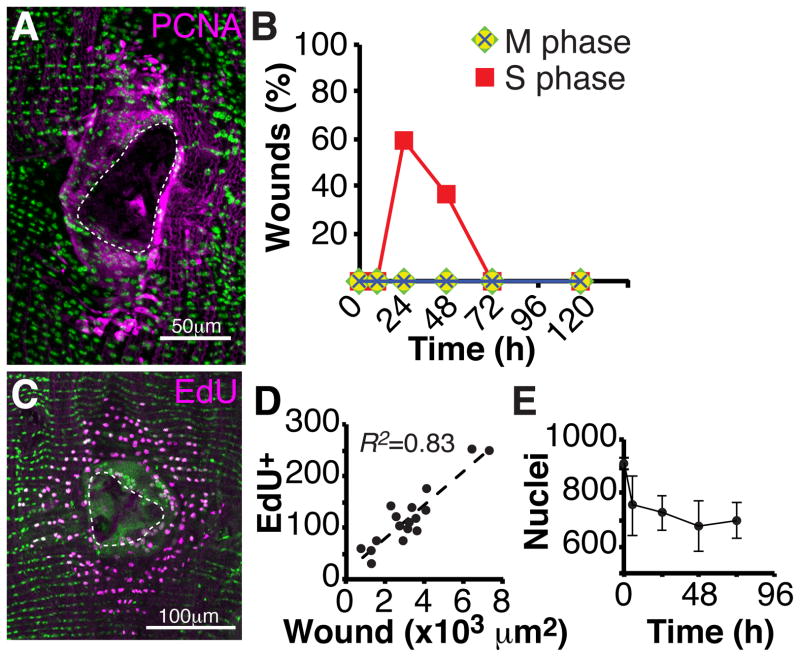
Conclusions: Our observations indicate that cell mass lost upon wounding can be replaced by polyploidization instead of mitotic proliferation. We propose that large cells generated by polyploidization or cell fusion are essential because they are better able than diploid cells to mechanically stabilize wounds, especially those containing permanent acellular structures, such as scar tissue.


Figures

Figure 1
Figure 1. Re-epithelialization occurs following abdominal injury
(A) Adult female flies were wounded on either side of the central sternites, between numbers 2–6 with a tungsten needle. (B) A melanin scab forms rapidly at the site of injury and persists after healing as a scar (shown 15 days post injury). (C) Time course of scab/scar formation (black) and re-epithelialization (green) (N=10–20 wounds/ time point). (D) Cross section model of ventral tissue organization. A continuous epithelial sheet (e, green) is in direct contact with outer cuticle (c, black), which is lined with small hairs. Lateral muscle fibers (m, red) lie below epithelium; organizing the tissue into alternating rows of muscle and epithelial cells. (E) Immunofluorescent images of abdominal tissue prior to and during re-epithelialization are shown below, with interpretive diagrams above. Epithelium (green, Epithelial-Gal4/ UAS-tubGFP). Muscle (red, Phalloidin). Scar (dashed white line). Scale bar= 50μm. See also Figure S1 and Figure S2.
Figure 2
Figure 2. Epithelial cells re-enter the cell cycle, but do not divide
(A) Immunofluorescent image of PCNA-GFP (magenta) expression at 24 hours post injury. DAPI (green); Scar (dashed white line). (B) S phase, but not M phase cell cycle markers are expressed post injury. Time course of the percentage of wound zones with cells expressing the indicated cell cycle markers. S phase marker (PCNA-GFP, red square) and M phase markers (PH3, green diamond; CycB-GFP, yellow circle; Polo kinase-GFP, blue cross). N=30 wounds/ time point. (C) Immunofluorescent image of EdU-positive nuclei (magenta) within the wound zone following continuous EdU labeling and analysis at 3 days post injury. Epithelial nuclei (green, flpout nlsGFP; Epithelial-Gal4/ UAS-Flp); Scar (dashed white line). (D) The number EdU-positive nuclei correlates with wound area (N=17). (E) Nuclei number is not restored following epithelial repair. Time course of the abdominal epithelial nuclear number within a fixed zone (7.5 × 104 μm2) following injury at the zone’s center (N=5, mean ± SD).
Figure 3
Figure 3. Cell fusion and increased cell size are induced by wounding
Immunofluorescent images of wound zones reveal epithelial organization: (A) Uninjured or (B) 3 days post injury. Cell-cell junctions (magenta, αFasIII). Epithelial nuclei (green, flpout nlsGFP; Epithelial-Gal4/ UAS-Flp). Giant syncytium (dashed yellow line), small syncytium (white arrow), enlarged cells (white arrowheads). (C) Average cell size (μm2) increases after injury. (N=28, mean ± SD). (D) Syncytial area (×103 μm2) versus nuclear number in the giant syncytium, or the small peripheral syncytia (inset, same units). See also Figure S3.
Figure 4
Figure 4. Polyploidization is induced by wounding
(A) Immunofluorescent images of wound zone showing location (white boxes) of particular nuclei shown stained with DAPI in panels to the right. A′, diploid nucleus distant from wound; A″, polyploid nucleus proximal to wound; A‴, cluster of polyploid nuclei from giant syncytium. Cell-cell junctions (red, αFasIII). Epithelial nuclei (green, flpout nlsGFP; Epithelial-Gal4/ UAS-Flp). DAPI (blue/ white, DNA). (B) Percentage of individual nuclei at indicated times post injury in the wound periphery (white and red bars) or cluster averages from the giant syncytium (blue bars) with DNA content in three ploidy classes (N=3 wounds, mean ± SD); blue bar (N=23 clusters, pooled from 3 and 7 day wounds). (C) Re-injury enhances polyploidy. A wound zone prior to injury (left panels; arrowheads = diploid nuclei) and after 5 wounds to the same site during a two-week period (right panels; arrowheads = highly polyploid nuclei). Markers are the same as in A.
Figure 5
Figure 5. Yki regulates polyploidization and cell fusion
(A–C) Immunofluorescent images of wound zones at 3 days post injury reveal polyploidization and cell fusion in control (no transgene) (A), yki knockdown (B), or in cycE knockdown (C) flies. Epithelial-Gal4 drove expression of the indicated transgene. Cell-cell junctions (magenta, αFasIII). Epithelial nuclei (green, αGrh). Giant syncytium (dashed yellow line). Scar (dashed white line). (D) Yki and CycE are required for S phase re-entry. Number of EdU-positive nuclei/ wound zone following continuous labeling and analysis at 3 days post injury in controls and flies expressing the indicated transgenes driven by Epithelial-Gal4 at 25°C (N=30, mean ± SD). (E) Same as D, except UAS-Hpo was driven in the presence of Gal80ts, and flies were shifted to 29°C prior to wounding (N=7, mean ± SD). (F) Yki and CycE are required for cells to polyploidize in response to injury. Percentage of nuclei from the indicated genotypes in the wound periphery at 7 days post injury with DNA content in three ploidy classes. Epithelial-Gal4 drove the indicated transgenes (N=3, mean ± SD). (**) T-test, p ≤ 0.01. (G) Knocking down yki enhances syncytium size. Giant syncytial area (×103 μm2) versus nuclear number at 3–4 days post injury. (H) Knocking down yki or cycE has little effect on wound closure. Indicated transgenes were expressed with Epithelial-Gal4, UAS-mCD8-RFP (N=10 wounds/ time point). mCD8-ChRFP was used to visualize wound closure. See also Figure S4.
Figure 6
Figure 6. Cell fusion and polyploidization are both required for wound closure
(AD) Immunofluorescent images of wound zones reveal affects of the indicated transgenes driven by Epithelial-Gal4 on cell fusion and wound healing at 3 days post injury. Cell-cell junctions (magenta, αFasIII). Epithelial nuclei (green, αGrh). Giant syncytium (dashed yellow line). (E) Epithelial expression of RacN17 reduces the size and nuclear content of the giant syncytium at 3–4 days post injury. (F) Blocking cell fusion alone or in combination with S phase inhibitory factors delays wound closure. Indicated transgenes were expressed with Epithelial-Gal4, UAS-mCD8-RFP. mCD8-ChRFP was used to visualize wound closure. (N=10–20 wounds/ time point). (G) Number of nuclei incorporating EdU following wounding of flies expressing the indicated transgenes as in F (N=7, mean ± SD). (H) Epithelial expression of RacN17 enhances polyploidization. Percentage of nuclei in the wound periphery from control (black bars, Epithelial-Gal4, no transgene) or Epithelial-Gal4, UAS-RacN17 flies at 7 days post injury with DNA content in three ploidy classes (N=3, mean ± SD). (*) T-test, p ≤ 0.05. (I) Models illustrating how blocking cell fusion (left) or fusion and polyploidization (right) affects wound closure. Only when both pathways are blocked do wounds remain open. The epithelial layer does not reseal under the scar. Scar is shown in dark brown. See also Figure S5.
Figure 7
Figure 7. Cell growth and polyploidy occur in response to hindgut tissue damage
(A) Scheme for genetic cell ablation in the adult hindgut. At 18°C, the Gal80ts repressor inhibits Gal4, preventing it from binding to UAS elements upstream of the pro-apoptotic genes hid and rpr. At 29°C, Gal80ts is inactivated, and Gal4 drives apoptotic gene expression specifically in the hindgut, under control of brachyenteron (byn) expression. (B–E) Changes in cell and nuclear size after injury, insets show magnified views of same image. (B) Cell-cell junctions and cell size in the undamaged adult pylorus are visualized with the adherens junction protein DE-Cadherin. (C) Two weeks after acute injury, cell-cell junction contacts appear intact around the now larger pyloric cells. (D) Nuclei in the undamaged adult pylorus. (E) Nuclei in the repairing adult pylorus. (F) Cell number does not recover in the adult pylorus after acute damage (control: N=6, mean ± SD; 7d: N=12, mean ± SD). (G) Measurements of nuclear ploidy in the adult pylorus (N=3, mean ± SD). Scale bars (white=25μm, red=12.5 μm).

References

    1. Razzell W, Wood W, Martin P. Swatting flies: modelling wound healing and inflammation in Drosophila. Dis Model Mech. 2011;4:569–574. - PMC - PubMed
    1. Galko MJ, Krasnow MA. Cellular and genetic analysis of wound healing in Drosophila larvae. PLoS Biol. 2004;2:E239. - PMC - PubMed
    1. Ramet M, Lanot R, Zachary D, Manfruelli P. JNK signaling pathway is required for efficient wound healing in Drosophila. Dev Biol. 2002;241:145–156. - PubMed
    1. Wang Z, Wilhelmsson C, Hyrsl P, Loof TG, Dobes P, Klupp M, Loseva O, Morgelin M, Ikle J, Cripps RM, et al. Pathogen entrapment by transglutaminase--a conserved early innate immune mechanism. PLoS Pathog. 2010;6:e1000763. - PMC - PubMed
    1. Tang H. Regulation and function of the melanization reaction in Drosophila. Fly (Austin) 2009;3:105–111. - PubMed

Publication types

MeSH terms