Review
. 2013 Dec;29(12):593-602.
doi: 10.1016/j.pt.2013.10.002. Epub 2013 Nov 1.

Regulation of immunopathogenesis during Plasmodium and Toxoplasma infections: more parallels than distinctions?

Affiliations
Review

Regulation of immunopathogenesis during Plasmodium and Toxoplasma infections: more parallels than distinctions?

Noah S Butler et al. Trends Parasitol. 2013 Dec.

Abstract

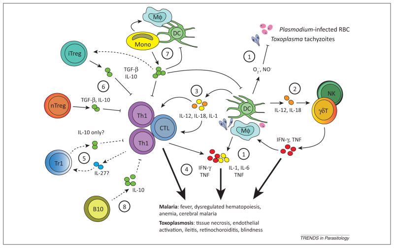
Toxoplasma and Plasmodium parasites exact a significant toll on public health. Host immunity required for efficient control of infection by these Apicomplexans involves the induction of potent T cell responses, which sometimes results in immunopathological damage. Thus, protective immune responses must be balanced by regulatory networks that limit immunopathology. We review several key cellular and molecular immunoregulatory networks operational during Toxoplasma and Plasmodium infections. Accumulating data show that despite differences in how the immune response controls these parasites, many host immunoregulatory pathways and cellular networks are common to both. Thus, understanding the cellular and molecular circuits that prevent or regulate immunopathological responses against one parasite is likely to inform our understanding of the host response to the other parasite.

Keywords: IL-10; IL-27; Plasmodium; TGF-β; Toxoplasma; immunopathology.


Figures

Figure 1
Figure 1
Common regulatory networks limit Toxoplasma and Plasmodium immunopathogenesis. (1) Recognition of parasites or parasite-infected cells by macrophages (Mφ) and dendritic cells (DCs) triggers the production of antiparasitic reactive oxygen species and synthesis of inflammatory cytokines [–92]. These inflammatory agents can act directly on parenchymal cells to induce necrotic or apoptotic pathways causing tissue damage, or (2) indirectly through the activation of innate natural killer (NK) or γδ T cells (γδT) [93,94]. (3) Cytokines such as IL-12, IL-1, and IL-18 also play a critical role in shaping the inflammatory nature of adaptive T cell subsets, which include IFN-γ secreting T helper cells (Th1) and cytotoxic cells (CTL) [85,93]. (4) Effector T cells express several inhibitory receptors including programmed death-1 (PD-1) and cytotoxic T lymphocyte antigen-4 (CTLA-4) that act to limit their activation, proliferation, and cytokine expression, which helps maintain immune homeostasis and limit immunopathology after infection [5,6,11,13]. Generally, and despite inhibitory receptor expression, during acute intestinal or ocular toxoplasmosis and malaria, highly activated T cells appear to directly contribute to pathology [37,46,98,104]. Pathological inflammatory reactions are themselves counterbalanced by several secreted regulatory cytokines and functionally distinct immune cell subsets. Regulatory cytokines such as IL-10 and TGF-β can be secreted by multiple cell types, including B cells, T cells, monocytes (Mono), Mφ, and DCs [2,17,18,24,26,29,51,67]. (5) Polarized IFN-γ-secreting Th1 cells also have the capacity to secrete IL-10 after their differentiation into Treg-like (Tr1) cells. Tr1 differentiation may involve IL-27 [23]. (6) Natural, thymus-derived Tregs (nTregs) suppress the activity of inflammatory CTL and Th1 cells through direct contact or secretion of IL-10 and TGF-β [12,46,106]. The precise role of nTreg, inducible Treg (iTreg), and Tr1 in limiting immunopathology during malaria and toxoplasmosis is not well understood. (7) Monocytes that infiltrate Toxoplasma-infected tissues or the spleen of Plasmodium-infected hosts exhibit dual roles in pathology and protection, by either secreting inflammatory cytokines (e.g., IL-12, TNF-α, IL-1, IL-6) or regulatory cytokines (IL-10 or TGF-β), and possibly by driving the differentiation of Tregs [–56]. The cues that determine whether monocytes exhibit an inflammatory or regulatory function are unknown. Additional questions include the specific role of IL-27 and the cellular source(s) of this cytokine, as well as (8) the contribution of IL-10-secreting regulatory B cells (B10). Well-described pathways are shown as unbroken lines, and unknown or speculative pathways are shown as broken lines.

References

    1. Stumhofer JS, et al. Interleukin 27 negatively regulates the development of interleukin 17-producing T helper cells during chronic inflammation of the central nervous system. Nat Immunol. 2006;7:937–945. - PubMed
    1. Gazzinelli RT, et al. In the absence of endogenous IL-10, mice acutely infected with Toxoplasma gondii succumb to a lethal immune response dependent on CD4+ T cells and accompanied by overproduction of IL-12, IFN-γ and TNF-α. J Immunol. 1996;157:798–805. - PubMed
    1. Wilson EH, et al. A critical role for IL-10 in limiting inflammation during toxoplasmic encephalitis. J Neuroimmunol. 2005;165:63–74. - PubMed
    1. Wherry EJ. T cell exhaustion. Nat Immunol. 2011;12:492–499. - PubMed
    1. Butler NS, et al. Therapeutic blockade of PD-L1 and LAG-3 rapidly clears established blood-stage Plasmodium infection. Nat Immunol. 2012;13:188–195. - PMC - PubMed

Publication types

Substances