. 2014 Jan;18(1):118-29.
doi: 10.1016/j.media.2013.10.001. Epub 2013 Oct 14.

Fully automatic segmentation of the mitral leaflets in 3D transesophageal echocardiographic images using multi-atlas joint label fusion and deformable medial modeling

Affiliations

Fully automatic segmentation of the mitral leaflets in 3D transesophageal echocardiographic images using multi-atlas joint label fusion and deformable medial modeling

A M Pouch et al. Med Image Anal. 2014 Jan.

Abstract

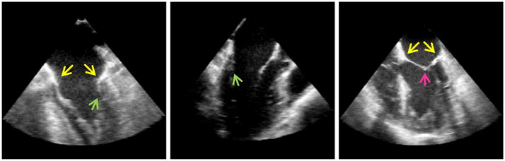
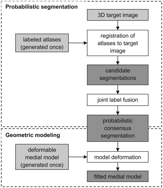
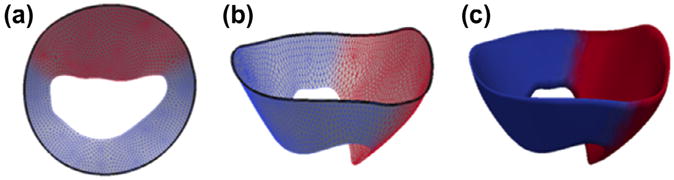
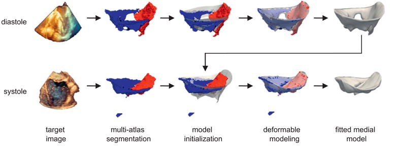
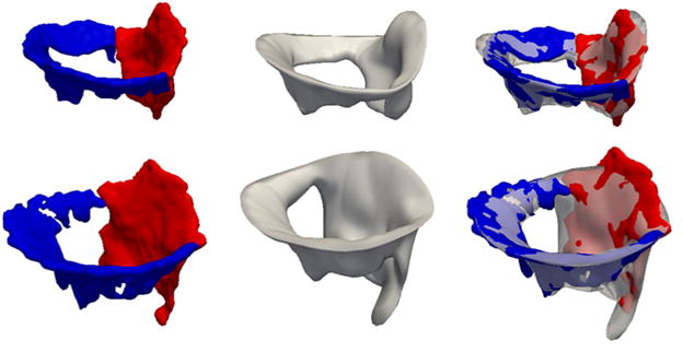
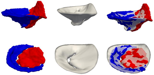
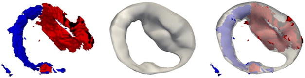
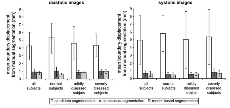
Comprehensive visual and quantitative analysis of in vivo human mitral valve morphology is central to the diagnosis and surgical treatment of mitral valve disease. Real-time 3D transesophageal echocardiography (3D TEE) is a practical, highly informative imaging modality for examining the mitral valve in a clinical setting. To facilitate visual and quantitative 3D TEE image analysis, we describe a fully automated method for segmenting the mitral leaflets in 3D TEE image data. The algorithm integrates complementary probabilistic segmentation and shape modeling techniques (multi-atlas joint label fusion and deformable modeling with continuous medial representation) to automatically generate 3D geometric models of the mitral leaflets from 3D TEE image data. These models are unique in that they establish a shape-based coordinate system on the valves of different subjects and represent the leaflets volumetrically, as structures with locally varying thickness. In this work, expert image analysis is the gold standard for evaluating automatic segmentation. Without any user interaction, we demonstrate that the automatic segmentation method accurately captures patient-specific leaflet geometry at both systole and diastole in 3D TEE data acquired from a mixed population of subjects with normal valve morphology and mitral valve disease.

Keywords: 3D echocardiography; Label fusion; Medial representation; Mitral valve; Multi-atlas segmentation.


Figures

Fig. 1
Fig. 1
Cross-sectional images of 3D TEE image volumes at diastole (left and center) and systole (right), illustrating the challenges specific to mitral leaflet segmentation. The yellow arrows point toward points on the annulus, showing that there is no image-based boundary between the mitral leaflets and adjacent tissue to which the leaflets are attached. The green arrows point towards the posterior leaflet at diastole, which is often pressed against the ventricular wall and is characterized by signal dropout. The pink arrow points to the coaptation zone of the leaflets at systole, showing there is no intensity-based demarcation between the anterior and posterior leaflets. (For interpretation of the references to color in this figure legend, the reader is referred to the web version of this article.)
Fig. 2
Fig. 2
Schematic of the automatic segmentation algorithm. The input is shown in light gray and the intermediate products and output are shown in dark gray. First, a set of 3D TEE atlases of the mitral leaflets is generated and a deformable medial model is constructed. Atlas and template generation is performed once. Given a 3D target image to segment, the atlases are registered to the target image and the atlas labels are propagated to the target image to obtain a set of candidate segmentations. Joint label fusion generates a probabilistic consensus segmentation, which is used to guide 3D deformable modeling. The output of the algorithm is a 3D geometric model of the mitral leaflets in the target image.
Fig. 3
Fig. 3
Cm-rep template of the open mitral leaflets used for deformable modeling. (a and b) The medial manifold of the template is a triangulated mesh, with red nodes referring to the anterior leaflet and blue labels referring to the posterior leaflet. The mitral annulus is represented by the bold black curve on the outer medial edge. (c) Leaflet boundaries constructed analytically from the medial manifold, given a constant radial thickness for initialization. (For interpretation of the references to color in this figure legend, the reader is referred to the web version of this article.)
Fig. 4
Fig. 4
Automatic segmentation of the mitral leaflets at diastole (top row) and systole (bottom row) for a given patient. First, a probabilistic segmentation is generated by multi-atlas label fusion (red = anterior leaflet, blue = posterior leaflet). Then the cm-rep template (translucent) is initialized to the multi-atlas segmentation and the template is deformed to obtain a medial model of the mitral leaflets. The medial template shown in Fig. 3 is used for model initialization at diastole, and the fitted diastolic model is used to initialize model fitting of the same subject's valve at systole. (For interpretation of the references to color in this figure legend, the reader is referred to the web version of this article.)
Fig. 5
Fig. 5
Automatic and manual open-leaflet segmentations for two subjects: one with a normal mitral valve (top row) and one with an incompetent valve (second row). The left column shows the manual segmentation with the anterior leaflet in red and posterior leaflet in blue. The center column shows the automatic segmentation, and the right column shows the automatic segmentation overlaid on the manual segmentation. (For interpretation of the references to color in this figure legend, the reader is referred to the web version of this article.)
Fig. 6
Fig. 6
Automatic and manual closed-leaflet segmentations for two subjects: one with a normal mitral valve (top row) and one with an incompetent valve (second row). The left column shows the manual segmentation with the anterior leaflet in red and posterior leaflet in blue. The center column shows the automatic segmentation, and the right column shows the automatic segmentation overlaid on the manual segmentation. (For interpretation of the references to color in this figure legend, the reader is referred to the web version of this article.)
Fig. 7
Fig. 7
The multi-atlas segmentation of an open valve, with the anterior leaflet in red and posterior leaflet in blue (left). The fitted cm-rep of the open leaflets (center). The fitted model overlaid on multi-atlas segmentation (right), illustrating that cm-rep corrects for labeling and topological inconsistencies in multi-atlas segmentation. (For interpretation of the references to color in this figure legend, the reader is referred to the web version of this article.)
Fig. 8
Fig. 8
Segmentation results at diastole (left) and systole (right). The bar graphs show the mean boundary displacement between the manual segmentation and each of the following: the candidate segmentations obtained by single-atlas registration, the consensus segmentation generated by joint label fusion, and the model-based segmentation created with the deformable cm-rep. The results are broken down by disease category: all subjects, subjects with normal mitral valve function, mildly diseased subjects, and subjects with severe mitral regurgitation.
Fig. 9
Fig. 9
Color maps showing localized mean distance between the automatic and manual segmentations in millimeters. The results are shown for the leaflets at systole (left column) and diastole (right column). Segmentation accuracy is evaluated in terms of a single-label model (top row) and multi-label model (bottom row), in which the anterior and posterior leaflet segmentations are evaluated independently. (For interpretation of the references to color in this figure legend, the reader is referred to the web version of this article.)

References

    1. Abraham TP, Warner JG, Jr, Kon ND, Lantz PE, Fowle KM, Brooker RF, Ge S, Nomeir AM, Kitzman DW. Feasibility, accuracy, and incremental value of intraoperative three-dimensional transesophageal echocardiography in valve surgery. Am J Cardiol. 1997;80:1577–1582. - PubMed
    1. Ahmed S, Nanda NC, Miller AP, Nekkanti R, Yousif AM, Pacifico AD, Kirklin JK, McGiffin DC. Usefulness of transesophageal three-dimensional echocardiography in the identification of individual segment/scallop prolapse of the mitral valve. Echocardiography. 2003;20:203–209. - PubMed
    1. Artaechevarria X, Munoz-Barrutia A, Ortiz-de-Solorzano C. Combination strategies in multi-atlas image segmentation: application to brain MR data. IEEE Trans Med Imaging. 2009;28:1266–1277. - PubMed
    1. Avants BB, Epstein CL, Grossman M, Gee JC. Symmetric diffeomorphic image registration with cross-correlation: evaluating automated labeling of elderly and neurodegenerative brain. Med Image Anal. 2008;12:26–41. - PMC - PubMed
    1. Avants BB, Tustison NJ, Song G, Cook PA, Klein A, Gee JC. A reproducible evaluation of ANTs similarity metric performance in brain image registration. Neuroimage. 2011;54:2033–2044. - PMC - PubMed

Publication types

MeSH terms