. 2014 Apr;35(4):772-7.
doi: 10.3174/ajnr.A3745. Epub 2013 Nov 1.

Craniopharyngeal canal and its spectrum of pathology

Affiliations

Craniopharyngeal canal and its spectrum of pathology

T A Abele et al. AJNR Am J Neuroradiol. 2014 Apr.

Abstract

Background and purpose: The craniopharyngeal canal is a rare, well-corticated defect through the midline of the sphenoid bone from the sellar floor to the anterosuperior nasopharyngeal roof. We reviewed a series of craniopharyngeal canals to determine a system of classification that might better our understanding of this entity, highlight the range of associated pathologic conditions, and optimize patient treatment.

Materials and methods: Available MR imaging, CT, and clinical data (from 1989-2013) of 29 patients (10 female, 15 male, 4 unknown; median age, 4 years; age range, 1 day-65 years) with craniopharyngeal canals were retrospectively examined. Qualitative assessment included orthotopic or ectopic adenohypophysis and the presence of a tumor and/or cephalocele. The midpoint anteroposterior diameter was measured. Clinical and imaging data were evaluated for pituitary dysfunction and accompanying anomalies.

Results: Craniopharyngeal canals were qualitatively separated into 3 types: incidental canals (type 1); canals with ectopic adenohypophysis (type 2); and canals containing cephaloceles (type 3A), tumors (type 3B), or both (type 3C), including pituitary adenoma, craniopharyngioma, dermoid, teratoma, and glioma. Quantitative evaluation showed a significant difference (P < .0001) in the anteroposterior diameters of type 1 canals (median, 0.8; range, 0.7-1.1 mm), type 2 canals (median, 3.9, range, 3.5-4.4 mm), and type 3 canals (median, 9.0; range, 5.9-31.0 mm) imparting small, medium, and large descriptors. Canals with cephaloceles all contained an ectopic adenohypophysis. The craniopharyngeal canals were associated with pituitary dysfunction (6/29) and congenital anomalies (8/29).

Conclusions: Accurate diagnosis and classification of craniopharyngeal canals are valuable to characterize lesions requiring surgery, identify patients with potential pituitary dysfunction, and avoid iatrogenic hypopituitarism or CSF leak during surgical resection of nasopharyngeal masses.


Figures

Fig 1.
Fig 1.
A 15-month-old girl with a history of trauma and type 1 CPC. A, Sagittal CT reconstruction shows the classic appearance of a small incidental CPC (arrow). Note its extent from the sella turcica to the roof of the nasopharynx. The spheno-occipital synchondrosis (dashed arrow) is visualized in its normal position posterior and inferior to the sella. B, Axial CT image shows well-corticated CPC (arrow) in the midline of the sphenoid body.
Fig 2.
Fig 2.
A 4-year-old boy with hypopituitarism and a type 2 CPC. A, Sagittal T1 MR image shows a medium-sized canal with ectopic, inferiorly displaced pituitary tissue (arrow). The AP diameter of this canal measured 3.5 mm. Note the normal spheno-occipital synchondrosis (dashed arrow). B, Coronal T1 MR image demonstrates soft tissue within the CPC (arrow). There is mild inferior displacement of the optic chiasm (dashed arrow) and infundibular recess of the third ventricle.
Fig 3.
Fig 3.
A 10-year-old boy with growth hormone deficiency and short stature with a type 3A CPC. A, Sagittal T1 MR image shows a large CPC containing a cephalocele with herniated adenohypophysis (arrow) and CSF. Note displacement of the infundibular recess of the third ventricle (dashed arrow). B, Sagittal T1 postcontrast MR image shows enhancement of the adenohypophysis (arrow), which, in our 4 type 3A canals, was invariably positioned along the posterior and/or inferior aspect of the cephalocele.
Fig 4.
Fig 4.
Two different patients with type 3 large CPCs containing tumor. A, Sagittal T1 MR image shows a large CPC containing portions of a T1 hyperintense dermoid (arrow) in a 30-year-old woman who presented with a nasopharyngeal mass. Note the associated cephalocele with herniated CSF and the adenohypophysis (dashed arrow). This is a type 3B CPC. B, Sagittal T1 postcontrast MR image in an 8-week-old girl who underwent imaging for a nasopharyngeal mass demonstrates a nasopharyngeal glioma (arrows), with mild enhancement extending into the suprasellar space through a large CPC (dashed arrows). This is a type 3C CPC.
Fig 5.
Fig 5.
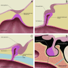
Embryology of the normal pituitary gland. A, At 3–4 weeks of gestation, the neuroectodermal adherence develops at a point of contact between the diencephalon (neurohypophyseal anlage) and roof of the stomodeum (adenohypophyseal anlage). B, At 4–5 weeks, the neuroectodermal adherence migrates dorsally forming the adenohypophyseal (Rathke) pouch, while the diencephalon migrates dorsal to the stomodeum to assume the position of the future neurohypophysis. The adenohypophyseal pouch elongates forming a stalk at its ventral aspect at 5–6 weeks (not shown). C, At 6–7 weeks, the postsphenoid cartilage develops, which results in obliteration of the adenohypophyseal stalk. D, In this illustration of the infant pituitary gland and sella, a dotted line traces the path of the potential CPC, which is believed to arise from nonobliteration of the adenohypophyseal stalk. The spheno-occipital synchondrosis (illustrated in blue) does not close until approximately age 16 years and mimics the CPC in children. Graphic illustrations used with permission from Amirsys.

References

    1. Kjaer I, Fischer-Hansen B. Human fetal pituitary gland in holoprosencephaly and anencephaly. J Craniofac Genet Dev Biol 1995;15:222–29 - PubMed
    1. Kaushik C, Ramakrishnaiah R, Angtuaco EJ. Ectopic pituitary adenoma in persistent craniopharyngeal canal: case report and literature review. J Comput Assist Tomogr 2010;34:612–14 - PubMed
    1. Hori A, Schmidt D, Rickels E. Pharyngeal pituitary: development, malformation, and tumorigenesis. Acta Neuropathol 1999;98:262–72 - PubMed
    1. Currarino G, Maravilla KR, Salyer KE. Transsphenoidal canal (large craniopharyngeal canal) and its pathologic implications. AJNR Am J Neuroradiol 1985;6:39–43 - PMC - PubMed
    1. Cohen R, Nelson MD, Segall HD. Epignathic teratoma associated with craniopharyngeal canal. AJNR Am J Neuroradiol 1989;10:652. - PMC - PubMed

MeSH terms