. 2013 Dec 1;191(11):5524-41.
doi: 10.4049/jimmunol.1102592. Epub 2013 Nov 1.

Brain human monoclonal autoantibody from sydenham chorea targets dopaminergic neurons in transgenic mice and signals dopamine D2 receptor: implications in human disease

Affiliations

Brain human monoclonal autoantibody from sydenham chorea targets dopaminergic neurons in transgenic mice and signals dopamine D2 receptor: implications in human disease

Carol J Cox et al. J Immunol. .

Abstract

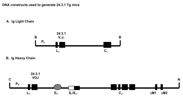
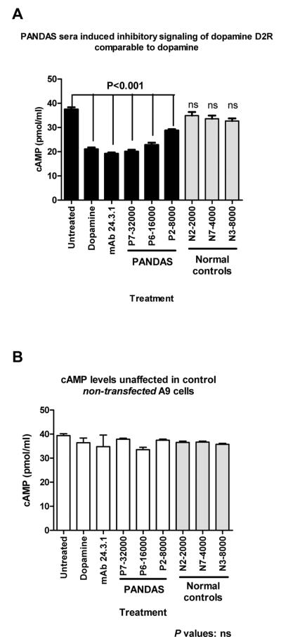
How autoantibodies target the brain and lead to disease in disorders such as Sydenham chorea (SC) is not known. SC is characterized by autoantibodies against the brain and is the main neurologic manifestation of streptococcal-induced rheumatic fever. Previously, our novel SC-derived mAb 24.3.1 was found to recognize streptococcal and brain Ags. To investigate in vivo targets of human mAb 24.3.1, VH/VL genes were expressed in B cells of transgenic (Tg) mice as functional chimeric human VH 24.3.1-mouse C-region IgG1(a) autoantibody. Chimeric human-mouse IgG1(a) autoantibody colocalized with tyrosine hydroxylase in the basal ganglia within dopaminergic neurons in vivo in VH 24.3.1 Tg mice. Both human mAb 24.3.1 and IgG1(a) in Tg sera were found to react with human dopamine D2 receptor (D2R). Reactivity of chorea-derived mAb 24.3.1 or SC IgG with D2R was confirmed by dose-dependent inhibitory signaling of D2R as a potential consequence of targeting dopaminergic neurons, reaction with surface-exposed FLAG epitope-tagged D2R, and blocking of Ab reactivity by an extracellular D2R peptide. IgG from SC and a related subset of streptococcal-associated behavioral disorders called "pediatric autoimmune neuropsychiatric disorder associated with streptococci" (PANDAS) with small choreiform movements reacted in ELISA with D2R. Reaction with FLAG-tagged D2R distinguished SC from PANDAS, whereas sera from both SC and PANDAS induced inhibitory signaling of D2R on transfected cells comparably to dopamine. In this study, we define a mechanism by which the brain may be altered by Ab in movement and behavioral disorders.


Figures

FIGURE 1
FIGURE 1. Schematic representation of DNA constructs used to generate 24.3.1 transgenic mice. A) Light chain construct
The Vκappa construct contains the human mAb 24.3.1 Sal I-flanked VJ region in addition to immunoglobulin promoter (Pκ) and leader (Lκ) sequences and the mouse κappa constant region (Cκ). B) Heavy chain construct. Human mAb 24.3.1 VDJ region flanked by Sal-I restriction sites was cloned into a mouse IgG1a transgenic construct previously described (Pogue and Goodnow). Restriction sites: B=BamHI; C=ClaI; N=NotI; S=SalI.
FIGURE 2
FIGURE 2. Expression of chimeric IgG1a in sera of transgenic (Tg) mice
Capture ELISA using anti-mouse IgG1a confirmed expression of 24.3.1 VH chimeric antibody in sera of 6-month-old 24.3.1 transgenic mice. (Dbl Tg=VH+VL)
FIGURE 3
FIGURE 3. Chimeric IgG1a transgenic antibody expressed in 24.3.1 Tg mouse sera and Tg-derived mAbs react with streptococcal and neuronal antigens
(A) Double (VH+VL) Tg mouse sera and (B) single (VH) Tg sera tested against a panel of antigens by ELISA using anti-mouse IgG1a retained cross-reactive antibody specificities to tubulin, lysoganglioside and streptococcal antigens. C) Antigen panel reactivity of 24.3.1 transgenic monoclonal antibodies (IgG1a) in the ELISA. Monoclonal Abs produced from Tg mouse splenic B cells retained cross-reactive specificities to tubulin, lysoganglioside and strep antigens. P-value was obtained by Tukey's test using one-way ANOVA; ns is not significant.
FIGURE 4
FIGURE 4. Chimeric Tg24.3.1 VH IgG1a Ab targeted dopaminergic neurons in Tg mouse brain in vivo
Colocalization of Tg 24.3.1 IgG1a (anti- IgG1a antibody-green) and tyrosine hydroxylase (TH) (anti-TH-antibody-red) by immunofluorescent staining of mouse brain tissue. TH is a marker for dopaminergic neurons. A–I) For each row, figure on left shows IgG1a (FITC-labeled), center shows tyrosine hydroxylase Ab (TRITC-labeled) and right figure is merged image (FITC-TRITC). A–C) Brain sections (basal ganglia) of Tg mouse (20X), showing FITC-labeled anti-mouse IgG1a (A), TRITC-labeled tyrosine hydroxylase Ab (B) and merged image (C). D–F) Enlarged images of A–C. G–I) Brain sections (basal ganglia) of non-Tg littermate tested with G) anti-mouse IgG1a, H) anti-TH Ab; I) merged image. J–K) Conjugated secondary antibody controls (secondary Ab, no primary Ab): J) FITC-conjugated streptavidin (1:20), K) TRITC-conjugated sheep anti-rabbit Ab (1:100). L) Tg mouse brain stained with DAPI. Outline denotes basal ganglia region of co-localization of images shown in Figures A–I. Shown at 4X. M–O) Cortex tissue section of Tg mouse brain: M) DAPI staining (10X) of cortex tissue section. Outline indicates location of 24.3.1 IgG1a positive (FITC-labeled) cells shown in Figure N; (O) Enlarged image of cortex tissue section. P–R) Hippocampus tissue section of Tg mouse brain: P) DAPI staining (10X) of hippocampus tissue section; Q) No staining for 24.3.1 IgG1a observed in the hippocampus; R) Enlarged image of hippocampus.
FIGURE 5
FIGURE 5. Tg24.3.1-IgG1a and human SC mAb 24.3.1 reacted and colocalized with dopamine receptor D2R. A) Reactivity of 24.3.1 Tg mAbs and Tg mouse serum (IgG1a) with human dopamine D2 receptor (D2R) in the ELISA using anti-mouse IgG1a
Tg mAbs reacted similar to human mAb 24.3.1 with D2R. Other human chorea-derived mAbs 37.2.1 and 31.1.1 reacted less with D2R than with mAb 24.3.1. Dbl Tg=VH+VL. P-value was obtained by Tukey's test using one-way ANOVA; ns is not significant. B) Chimeric Tg24.3.1 VH IgG1a Ab colocalized with anti-D2R in Tg mouse brain in vivo. Colocalization of Tg 24.3.1 IgG1a (anti- IgG1a antibody) and rabbit polyclonal anti-D2R Ab by immunofluorescent staining of mouse brain tissue. Top row: Brain sections of Tg mouse (20X), showing FITC-labeled anti-mouse IgG1a (left), TRITC-labeled anti-D2R Ab (center) and merged image (right). Middle row: Enlarged images of top row. Bottom row: Secondary antibody controls: FITC-conjugated streptavidin (1:20) (left), and TRITC-conjugated sheep anti-rabbit Ab (1:100) (right) were negative. C) Human SC-derived mAb24.3.1 (IgM) colocalized with anti-D2R in normal mouse brain tissue sections. Colocalization of human SC mAb 24.3.1 (anti-human IgM antibody) and rabbit polyclonal anti-D2R antibody in normal mouse brain tissue sections. Top row: Brain tissue sections of normal mouse (10X), incubated in vitro with human SC mAb 24.3.1, showing FITC-labeled anti-human IgM (left), TRITC-labeled rabbit polyclonal D2R Ab (center), and merged image (right). Second row: Enlarged images of top row. Media controls (IMDM supplemented with 10% FBS) and secondary antibody controls (FITC-conjugated anti-human IgM, 1:50; TRITC-conjugated sheep anti-rabbit Ab, 1:100) were negative. Concentrations of mAbs tested (24.3.1, 37.2.1, 31.1.1): 100 ng/ml.
FIGURE 6
FIGURE 6. Human SC mAb 24.3.1 reacted with FLAG epitope-tagged D2R at the cell surface and with a D2R peptide
A) Quantification of the relative levels of cell-surface binding of anti-FLAG M2 Ab to HEK-293T cells transfected with cDNA for the FLAG-tagged D2R construct or HEK-293T cells transfected with an empty vector (untransfected cells). White bars represent HEK-293T cells treated with control media (no antibody) and closed black bars represent HEK-293T cells treated with anti-FLAG M2 Ab (mean ± SD, n=5). B) Quantification of the relative levels of cell-surface binding of human chorea-derived SC mAbs to HEK-293T cells transfected with cDNA for the FLAG-tagged D2R construct (closed black bars) or with empty vector (untransfected cells, white bars) (mean ± SD, n=5). All the chorea-derived SC mAbs showed significant binding to D2R-transfectants compared to cells transfected with empty vector. C) Human SC sera reacted with FLAG epitope-tagged D2R at the cell surface. Quantification of the relative levels of cell-surface binding of human PANDAS sera (P), SC sera (SC), and sera from normal healthy controls (N) to HEK-293T cells transfected with cDNA for the FLAG-tagged D2R construct (closed black bars) or with empty vector (untransfected cells, grey bars) (mean ± SD, n= 4). Four SC sera tested (SC1-4) showed significant (p<0.001 or p<0.05) binding to D2R-transfectants compared to cells transfected with empty vector. Four PANDAS sera tested (P1–4) were negative and did not show significant binding. Normal control sera tested were negative. D) D2R peptide E1.1 significantly inhibited binding of human SC mAb 24.3.1 to dopamine D2 receptor in a dose-dependent manner. Figure 6D left shows dose-dependent inhibition of anti-D2R mAb 1B11 binding to D2R by extracellular D2R peptide E1.1 (p<0.001). Figure 6D right shows significant (p<0.001) inhibition of chorea-derived mAb 24.3.1 binding to D2R by D2R peptide E1.1. Control peptide and extracellular D2R peptide E1.2 did not strongly inhibit and behaved similarly when reacted with the commercial mouse anti-D2R mAb and our chorea-derived human mAb 24.3.1 as shown on right. P<0.001 (D2R E1.1 vs. E1.2). Media control (negative) not shown. E) Dose response inhibition of two other human SC mAbs 31.1.1 and 37.2.1 by D2R peptide E1.1. Figure 6E shows dose-dependent binding inhibition of human SC mAbs 31.1.1 and 37.2.1 to D2R by extracellular D2R peptide E1.1 (p<0.001). D2R peptide E1.2 did not show significant inhibition compared to E1.1 (mAb 31.1.1, p<0.001; mAb 37.2.1, p<0.01). D2R peptide E1.2 did not strongly inhibit. Extracellular D2R peptide E1.2 did not strongly inhibit. Media control (negative) not shown. mAb concentration: 100 ng/ml. F) SC Tg VH24.3.1 mAbs reacted with D2R peptides in the ELISA. Tg mAbs expressing human mAb 24.3.1 heavy chain (VH) were tested in the direct ELISA for reactivity with D2R peptides E1.1 and E1.2. Tg mAbs showed specificity for peptide D2R E1.1 (D2R E1.1 vs. D2R E1.2, p<0.001). Media control (negative) not shown.
FIGURE 7
FIGURE 7. IgG in SC and PANDAS reacted with dopamine receptor D2R. A) IgG from SC and PANDAS sera reacted with human D2R
Human sera from SC, PANDAS, Tourettes/OCD and ADHD were titrated for reactivity with human D2R. A strong reaction of SC (p=0.0005) and PANDAS sera (p=0.0291) with D2R in the ELISA is shown in Figure 7A. B, C) IgG from SC and PANDAS reacted with D2R compared to the different response against D1R. Reactivity of human sera from SC, PANDAS, Tourettes/OCD and ADHD with dopamine receptors D1R and D2R in the ELISA is shown. For PANDAS, p<0.0001 (D1R vs. D2R). For SC, p=0.001. Ab titers are shown in both graphs in Figure 7B and OD values are shown in Figure 7C for comparison of titers vs OD @ 405 (@1:500 dilution). P-values were obtained by Mann-Whitney and Kruskal-Wallis.
FIGURE 8
FIGURE 8. Human mAb 24.3.1 signaled human dopamine receptor D2 in transfected D2R cells by inhibiting adenylate cyclase activity comparable to dopamine
Inhibition of adenylate cyclase by receptor activation in human D2R-transfected cell lines (A9-hD2R) was quantified by cAMP levels measured in cell lysates. Incubation of the D2R cell line with chorea-derived mAbs 24.3.1, 31.1.1 and 37.2.1. Results are shown from cAMP direct competition immunoassay. A) D2R transfected cells treated with mAb 24.3.1 showed a 53% decrease in cAMP which was comparable to dopamine-treated cells (p<0.001). Cells treated with pertussis toxin are also shown; this treatment was used as an experimental control, since D2 receptor-mediated activation and inhibitory signaling with concomitant decreases in cAMP can be blocked by pre-incubation of cells with pertussis toxin. B) Unresponsive control (non-transfected) A9 cells treated with dopamine and mAbs. C) Dose-response curve showed cAMP levels in D2R transfected cells following treatment with different concentrations of mAb 24.3.1 at 1, 5, 10, 25 and 50 ngs, and unresponsive control (non-transfected) A9 cells after same treatment. Assays were performed in triplicate. P-value was obtained by Tukey's test using one-way ANOVA; ns is not significant. Concentrations of mAbs tested are as follows: 24.3.1 ~90 ng/ml; 31.1.1 ~100 ng/ml; 37.2.1 ~100 ng/ml.
FIGURE 9
FIGURE 9. D2R transfectants treated with human SC sera with elevated anti-D2R antibody show decreased cAMP levels
Inhibition of adenylate cyclase by receptor stimulation in human D2R-transfected cell lines (A9-hD2R) was quantified by cAMP levels measured in cell lysates. Incubation with human SC mAb 24.3.1 antibody donor serum, other SC sera, and normal healthy control sera. Results are shown from cAMP direct competition immunoassay. A) D2R transfected cells treated with SC antibody donor serum (SC6; anti-D2R titer 32,000) and SC serum 7 (SC7, anti-D2R titer 16,000) showed 40% decrease in cAMP levels comparable to dopamine (48%) and mAb 24.3.1-treated cells (43% decrease); p<0.0001. cAMP levels were also significantly decreased (p<0.001) in transfectants treated with five other SC sera (SC1, SC2, SC3, SC5, SC8) with anti-D2R titers of 8,000 and 16,000. cAMP level was not significantly reduced in transfectants treated with normal serum (anti-D2R titers 2,000, 8,000 or 16,000). B) Control (non-transfected) A9 cells were unresponsive. Assays were performed in triplicate. P-value was obtained by Tukey's test using one-way ANOVA; ns: not significant. Concentration of mAb 24.3.1 ~90 ng/ml.
FIGURE 10
FIGURE 10. D2R transfected cells treated with PANDAS sera showed decreased cAMP levels compared to transfectants treated with normal control sera
A) D2R transfected cells were treated with PANDAS sera and sera from normal controls. PANDAS sera with elevated human anti-D2R Ab ELISA titers were assayed for inhibitory signaling [anti-D2R titers: P7 – 32,000; P6 – 16,000; P2 – 8,000] showed significantly decreased cAMP levels (46%, 39%, 23%) comparable to decreased cAMP levels in response to the neurotransmitter dopamine (44%) and chorea mAb 24.3.1 (48%) treatments. D2R transfectants treated with normal sera of various ELISA anti-D2R titers (N2 – 2,000, N7 – 4,000, N3 – 8,000) did not show significant reductions in cAMP. B) Control (non-transfected) A9 cells were unresponsive. Assays were performed in triplicate. P-value was obtained by Tukey's test using one-way ANOVA; ns: not significant. Concentration of mAb 24.3.1 ~90 ng/ml. PANDAS sera and normal controls: N=7 (three of each are shown in the figure).

References

    1. Stollerman GH. Rheumatic fever in the 21st century. Clin Infect Dis. 2001;33:806–814. - PubMed
    1. Marques-Dias MJ, Mercadante MT, Tucker D, Lombroso P. Sydenham's chorea. Psychiatr Clin North Am. 1997;20:809–820. - PubMed
    1. Taranta A, Stollerman GH. The relationship of Sydenham's chorea to infection with group A streptococci. Am J Med. 1956;20:170–175. - PubMed
    1. Taranta A. Relation of isolated recurrences of Sydenham's chorea to preceding streptococcal infections. N Engl J Med. 1959;260:1204–1210. - PubMed
    1. Kurlan R, Kaplan EL. The pediatric autoimmune neuropsychiatric disorders associated with streptococcal infection (PANDAS) etiology for tics and obsessive-compulsive symptoms: hypothesis or entity? Practical considerations for the clinician. Pediatrics. 2004;113:883–886. - PubMed

Publication types

MeSH terms