Review
. 2014 Jan 13:1542:206-20.
doi: 10.1016/j.brainres.2013.10.049. Epub 2013 Nov 1.

TAM receptor tyrosine kinases: expression, disease and oncogenesis in the central nervous system

Affiliations
Review

TAM receptor tyrosine kinases: expression, disease and oncogenesis in the central nervous system

Angela M Pierce et al. Brain Res. .

Abstract

Receptor tyrosine kinases (RTKs) are cell surface proteins that tightly regulate a variety of downstream intra-cellular processes; ligand-receptor interactions result in cascades of signaling events leading to growth, proliferation, differentiation and migration. There are 58 described RTKs, which are further categorized into 20 different RTK families. When dysregulated or overexpressed, these RTKs are implicated in disordered growth, development, and oncogenesis. The TAM family of RTKs, consisting of Tyro3, Axl, and MerTK, is prominently expressed during the development and function of the central nervous system (CNS). Aberrant expression and dysregulated activation of TAM family members has been demonstrated in a variety of CNS-related disorders and diseases, including the most common but least treatable brain cancer in children and adults: glioblastoma multiforme.

Keywords: Alzheimer's disease; Cell signaling; Developmental neurology; Glioblastoma; Multiple sclerosis.


Figures

Fig. 1
Fig. 1
TAM expression in the human brain. While Tyro3 is highly expressed in many regions of the adult human brain (Lemke and Rothlin, 2008; Mark et al., 1994), Axl and MerTK are present in fewer cell types and at lower levels. By immunohistochemical analysis MerTK is expressed in less than 5% of the astrocytes (arrowheads; top middle panel) while Axl expression is limited to the endothelial cells of the vasculature (arrowheads; bottom middle panel). Immunohistochemical staining of fixed adult human normal cerebrum was performed on normal brain obtained from University of Colorado Hospital. Paraffin embedded tissue blocks were cut into 5 um sections, deparaffinized in xylene, and rehydrated through a descending concentration sequence of ethanol. Sections were subjected to antigen retrieval in citrate buffer (pH 6.0) for 10 min in a decloaking chamber. Endogenous peroxidase activity and nonspecific binding was blocked by 3% hydrogen peroxide block for 15 min and then 3% BSA protein block for 15 min. Immunohistochemical staining was performed using labeled streptavidin biotin reagents (LSAB) on a Dako Autostainer (Dako, Carpinteria, CA). The primary antibodies, 0.2 ug/mL rabbit anti-MerTK antibody (Epitomics, Burlingame, CA) and 0.5 ug/mL goat anti-human Axl antibody (R&D Systems, Minneapolis, MN), were incubated at room temperature for 60 min, followed by the biotinlyated secondary antibody (LSAB+kit, Dako) for 30 min and streptavidin-horseradish peroxidase (LSAB+ kit, Dako) for 30 min. Localization of staining was performed with 3,3′ diaminobenzidine (Dako). Slides were counterstained with hematoxylin (Dako) and coverslipped using a xylene based mounting medium. Normal goat IgG (Santa Cruz Biotechnology, Santa Cruz, CA) and Rabbit Immunoglobin (Dako) were used in place of the primary antibodies for negative staining controls. Human Mer/Fc Chimera (R&D Systems) and Human Axl/Fc Chimera (R&D Systems) were preincubated with the respective primary antibodies, in five-fold excess by weight, to determine specificity of antibody binding. Magnification bars provided represent 10 um.
Fig. 2
Fig. 2
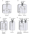
TAM signaling in cells of the CNS. The schematic represents a generalization of the important TAM signaling pathways within the CNS derived from a variety of studies. In many instances one or more receptors, or interactions between receptors, are responsible for the signaling pathways noted; this is a simplified summary representation. Refer to citations for specific details. (A) Protein S mediates survival of cortical and hippocampal neurons through Tyro3, regulating both the intrinsic and the extrinsic apoptotic pathways (Zhong et al., 2010). (B) GnRH NLT neurons, isolated from embryonic mouse brain, express no Tyro3 but abundant Axl and MerTK. Gas6 is the sole ligand recognized for Axl and stimulates both migration and survival through Axl in GnRH NLT cells (Allen et al., 1999; Nielsen-Preiss et al., 2007). (C) Gas6 does play a role in neuronal repair and synaptic plasticity: it binds Tyro3 in cortical and hippocampal neurons and upregulates components of transcriptional and translational machinery (Prieto et al., 2007). (D) Glial cells generally express much lower levels of Tyro3 than (adult) neurons. While oligodendroctyes express all three TAMs, Axl (not Tyro3) mediates survival signaling via Gas6, perhaps because Axl has greater affinity for Gas6 than Tyro3 does (Shankar et al., 2003; Weinger et al., 2008). (E) Microglia express Axl and MerTK but have no detectable levels of Tyro3. Gas6 has been shown to bind both Axl and MerTK, limiting inflammatory responses and promoting phagocytosis in microglia, while Tubby reportedly binds MerTK and promotes phagocytosis in macrophages and retinal epithelial pigment cells (Caberoy et al., 2009; Caberoy and Zhou, 2010).

Similar articles

Cited by

References

    1. Allen MP, et al. Growth arrest-specific gene 6 (Gas6)/adhesion related kinase (Ark) signaling promotes gonadotropin-releasing hormone neuronal survival via extracellular signal-regulated kinase (ERK) and Akt. Mol Endocrinol. 1999;13:191–201. - PubMed
    1. Anwar AA, et al. Mer tyrosine kinase (MerTK) promotes macrophage survival following exposure to oxidative stress. J Leukoc Biol. 2009;86:73–79. - PMC - PubMed
    1. Bellosta P, et al. The receptor tyrosine kinase ARK mediates cell aggregation by homophilic binding. Mol Cell Biol. 1995;15:614–625. - PMC - PubMed
    1. Bellosta P, et al. Signaling through the ARK tyrosine kinase receptor protects from apoptosis in the absence of growth stimulation. Oncogene. 1997;15:2387–2397. - PubMed
    1. Biesecker LG, Gottschalk LR, Emerson SG. Identification of four murine cDNAs encoding putative protein kinases from primitive embryonic stem cells differentiated in vitro. Proc Natl Acad Sci USA. 1993;90:7044–7048. - PMC - PubMed

MeSH terms

Substances