. 2014 Jan 23;123(4):509-19.
doi: 10.1182/blood-2013-07-515874. Epub 2013 Nov 1.

Thrombopoietin promotes NHEJ DNA repair in hematopoietic stem cells through specific activation of Erk and NF-κB pathways and their target, IEX-1

Affiliations

Thrombopoietin promotes NHEJ DNA repair in hematopoietic stem cells through specific activation of Erk and NF-κB pathways and their target, IEX-1

Bérengère de Laval et al. Blood. .

Abstract

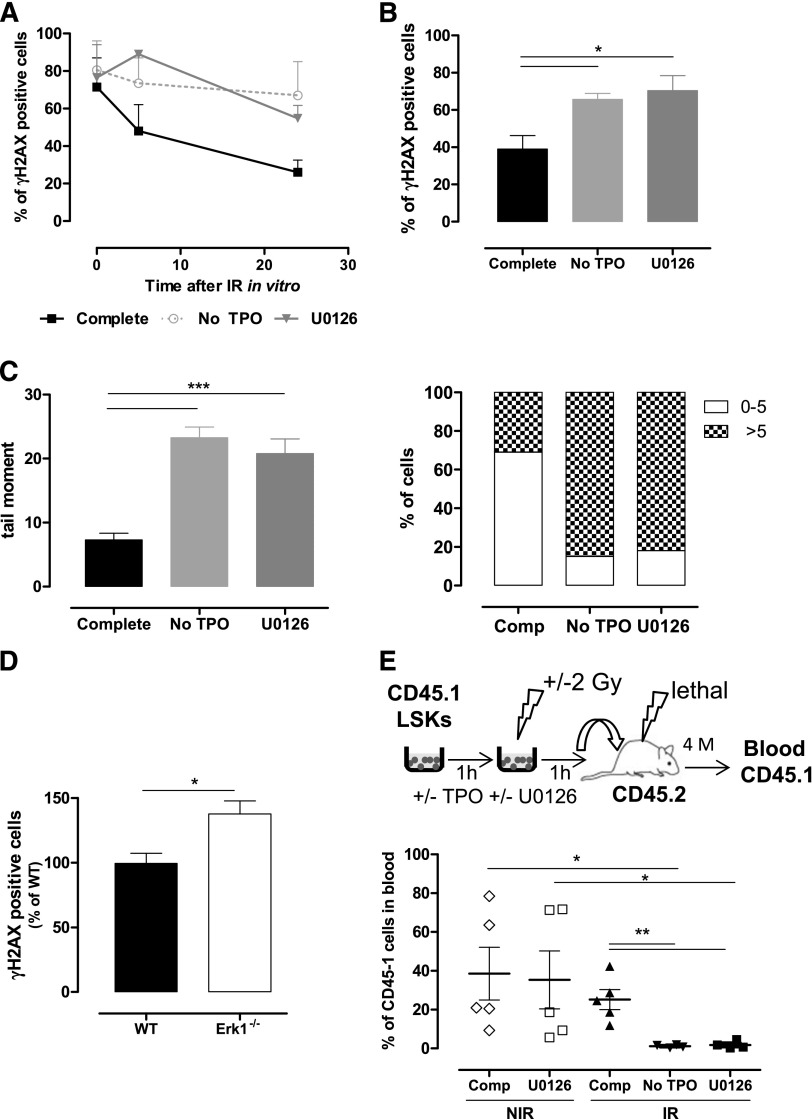
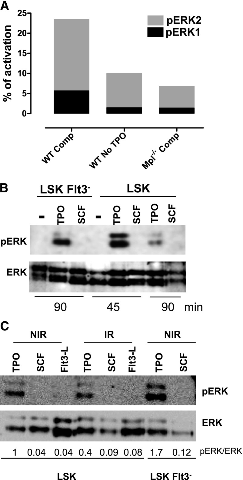
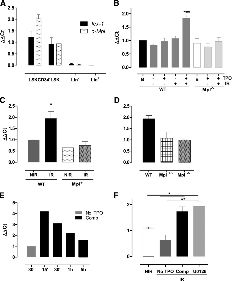
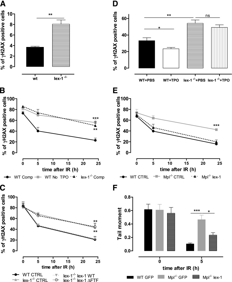
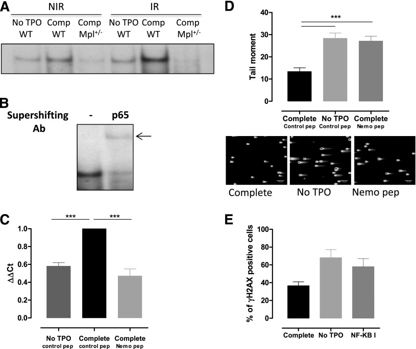
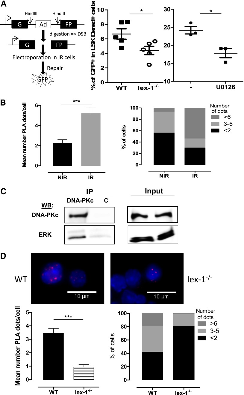
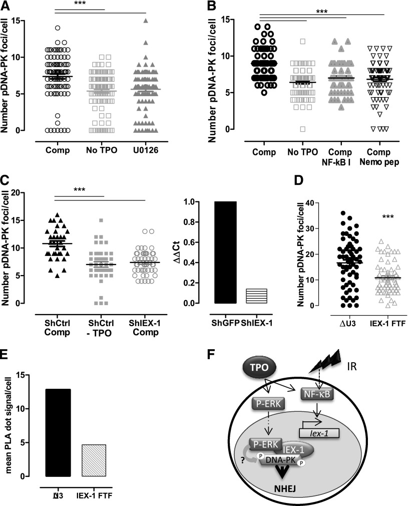
Loss of hematopoietic stem cell (HSC) function and increased risk of developing hematopoietic malignancies are severe and concerning complications of anticancer radiotherapy and chemotherapy. We have previously shown that thrombopoietin (TPO), a critical HSC regulator, ensures HSC chromosomal integrity and function in response to γ-irradiation by regulating their DNA-damage response. TPO directly affects the double-strand break (DSB) repair machinery through increased DNA-protein kinase (DNA-PK) phosphorylation and nonhomologous end-joining (NHEJ) repair efficiency and fidelity. This effect is not shared by other HSC growth factors, suggesting that TPO triggers a specific signal in HSCs facilitating DNA-PK activation upon DNA damage. The discovery of these unique signaling pathways will provide a means of enhancing TPO-desirable effects on HSCs and improving the safety of anticancer DNA agents. We show here that TPO specifically triggers Erk and nuclear factor κB (NF-κB) pathways in mouse hematopoietic stem and progenitor cells (HSPCs). Both of these pathways are required for a TPO-mediated increase in DSB repair. They cooperate to induce and activate the early stress-response gene, Iex-1 (ier3), upon DNA damage. Iex-1 forms a complex with pERK and the catalytic subunit of DNA-PK, which is necessary and sufficient to promote TPO-increased DNA-PK activation and NHEJ DSB repair in both mouse and human HSPCs.


Figures

Figure 1
Figure 1
Erk activation is necessary for TPO-mediated DSB repair in HSPCs. (A-B) γH2AX foci resolution analysis at the indicated times after IR (2 Gy) of (A) LSK and (B) LSK-CD34 cells cultured in complete medium in the presence or absence of the MEK inhibitor U0126 (10 µM), or in medium without TPO. Data are means + standard error of the mean (SEM) (n = 3). (C) DSB analysis using a neutral comet assay of LSK cells cultured as in panel A. Mean of tail moments (left) and percent of cells with tail moments >5 or <5 (right) are shown. Representative experiment out of 3 similar performed with cells pooled from 3 to 4 mice. Data are means + SEM of tail moment values analyzed in at least 100 cells. (D) γH2AX staining in LSK-CD34 cells isolated 16 hours after TBI of wild type (WT) and Erk1−/− mice. Data are means + SEM normalized to the mean of γH2AX positive cells from WT mice (n = 5). (E) Experimental design to test the effect of Erk inhibition on LSK reconstitution ability (top). LSK cells were cultured as in panel A for 1 hour before injection in CD45-2 lethally irradiated congenic mice. CD45.1-chimerism in peripheral blood 4 months posttransplantation (bottom). Each dot represents an individual mouse. Mice were injected with LSK cells from a pool of 9 mice. Comp, complete medium; NIR, no IR.
Figure 2
Figure 2
TPO is the major Erk activator in HSPCs. (A) One representative experiment, out of 3 similar experiments, of Erk activity obtained by capillary isoelectric-focusing immunoassay in WT and Mpl−/− LSK cells cultured for 1 hour in complete medium (Comp) or without TPO. Results show the area peak value of phosphorylated Erk1 or Erk2, normalized to the area value of total Erk. Experiment performed with cells pooled from 3 to 5 mice. (B-C) Western blot analysis of Erk activation in LSK or LSK-Flt3 cells cultured for 45 or 90 minutes as indicated with SCF, FL, or TPO or in the absence of cytokine (−) and then irradiated (2 Gy) or not. Analysis was performed 30 minutes after IR. NIR, no IR.
Figure 3
Figure 3
Iex-1 is induced in HSPCs in response to TPO and IR. (A) Quantitative polymerase chain reaction analysis of Mpl and Iex-1 mRNA levels in different hematopoietic cell populations. Results are means + SEM (n = 3). (B-F) Quantitative polymerase chain reaction analysis of Iex-1 expression in LSK cells from WT, Mpl−/−, and Mpl+/− mice cultured in vitro in complete medium (Comp) or without TPO and irradiated or not (2 Gy). Analysis was performed at 5 hours (B,D) or at various times after IR (E). (F) Cells were incubated with the MEK inhibitor U0126 (10 µM) 30 minutes before being treated as in panel B. Data are means + SEM (n = 4). (C) Iex-1 mRNA expression in LSK cells isolated 5 hours after TBI of WT and Mpl−/− mice. Data are means + SEM (n = 3). All the results are normalized on gapdh expression.
Figure 4
Figure 4
Iex-1 is necessary and sufficient for TPO-mediated DSB repair in HSPCs. (A) Percentage of γH2AX-positive LSK cells isolated from nontreated WT and Iex-1−/− mice. Data are means + SEM (n = 3). (B) Kinetics of γH2AX foci resolution after IR (2 Gy) of WT and Iex-1−/− LSK cells cultured in complete medium (Comp) or without TPO. (C) WT LSK cells were infected with lentiviruses expressing GFP only or GFP with Iex-1-WT or Iex-1-ΔFTF and treated as in (B). For (B) and (C), the results represent means + SEM of 3 independent experiments performed with pools of 5 to 10 mice. (D) WT and Iex-1−/− mice were treated with TPO (8 µg/kg) or phosphate-buffered saline 30 minutes before TBI (2 Gy). γH2AX staining in LSK-CD34 cells isolated 16 hours after TBI. (E-F) γH2AX foci (E) and comet tail resolution (F) after IR of WT and Mpl−/− LSK cells infected as in (C) and cultured in the presence of TPO complete medium. Means + SEM are shown. (E) n = 4. (F) Data are means + SEM of tail moment values analyzed in at least 35 cells.
Figure 5
Figure 5
NF-κB is required for TPO-induced Iex-1 expression and DSB repair in HSPCs. (A) EMSA for NF-κB DNA-binding activity in WT and Mpl+/− LSK cells cultured in complete medium (Comp) or without TPO for 45 minutes and then irradiated (2 Gy) or not. Analysis was performed 30 minutes after IR. Representative experiment out of 4 similar performed. (B) Supershift analysis after addition of anti-p65RelA antibody or no antibody (−) in WT LSK cells treated as in (B) 30 minutes post-IR in the presence of TPO. (C) Iex-1 mRNA expression 30 min after IR in LSK cells precultured in medium without TPO or in complete medium containing either the Nemo peptide or a control peptide (10 µM). Data are means + SEM from 4 independent experiments performed with cells from 3 to 5 mice. (D) Comet tail moment of WT LSK cultured as in panel C and 2 hours after IR. Representative pictures of comets labeled with SYTOXGreen and means + SEM of the tail moment values measured in at least 60 cells are shown. The scale bars on the pictures represent a 107 µm × 10 objective. One representative experiment out of 3 experiments is shown. (E) γH2AX staining in LSK cells cultured like in panel D in the presence or absence or of the NF-κB inhibitor (NF-κB I, 1 µM) 24 hours after IR. Data are means + SEM of 3 independent experiments with at least 100 cells counted per condition.
Figure 6
Figure 6
pErk and DNA-PKc form a complex that requires the presence IEX-1. (A) NHEJ activity in WT and Iex-1−/− LSK cells cultured in complete medium in the presence or absence of U0126 (10 μM). The experimental procedure (left) and means + SEM percent of GFP+ cells in LSK DsRed+ cells (right) are shown. Each dot represents an individual mouse. (B,D) PLA between pErk and DNA-PKc in WT and Iex-1−/− LSK cells. Cells were cultured in complete medium for 30 minutes and analyzed 30 minutes after IR (2 Gy) or not. Representative pictures (×100) and means + SEM of PLA dot numbers per cell (left) and percent of cells with the indicated amounts of dots (right) are shown. (C) UT7-Mpl cells were treated with TPO and then irradiated (2 Gy). Thirty minutes after IR, nuclear extracts were immunoprecipitated with anti-DNA-PKc or control (c) mouse immunoglobulin G. NIR, no IR.
Figure 7
Figure 7
Erk and IEX-1 are necessary for DNA-PK activation in human and mouse HSPCs. (A-B) Ser2056-pDNA-PK foci number 30 minutes after IR of CD34+ human progenitor cells cultured without TPO or in complete medium (Comp) in the presence or absence of 10 µM U0126 (A) or 1 µM NF-κB I or 10 µM Nemo inhibitory peptide (B). Each dot represents an individual cell. Data are means + SEM from cells counted in 3 independent experiments. (C) Number of pSer2056-pDNA-PK 30 minutes after IR of CD34+ human cells infected with lentiviruses encoding control GFP-shRNA or IEX-1-shRNA and cultured and analyzed as in panel A (left). Data are means + SEM foci per cell counted from 3 independent experiments. IEX-1 mRNA expression in the shRNA-infected cells analyzed on the left (right). (D) Number of Thr2609-pDNA-PK and (E) pErk/DNA-PKc interaction using PLA in UT7-Mpl infected with control (ΔU3) or with IEX-1-ΔFTF vectors and treated with TPO and IR as in panel A. Representative experiments. (F) Representative model illustrating the cooperation between NF-κB and Erk activation upon TPO and IR, leading to Iex-1 induction and Iex-1 activation, respectively. The complex formed between pErk/Iex-1/DNA-PKc is required for DNA-PK phosphorylation and DNA-repair promotion.

Comment in

References

    1. Simonnet AJ, Nehmé J, Vaigot P, Barroca V, Leboulch P, Tronik-Le Roux D. Phenotypic and functional changes induced in hematopoietic stem/progenitor cells after gamma-ray radiation exposure. Stem Cells. 2009;27(6):1400–1409. - PubMed
    1. Wang Y, Liu L, Pazhanisamy SK, Li H, Meng A, Zhou D. Total body irradiation causes residual bone marrow injury by induction of persistent oxidative stress in murine hematopoietic stem cells. Free Radic Biol Med. 2010;48(2):348–356. - PMC - PubMed
    1. Fleenor CJ, Marusyk A, DeGregori J. Ionizing radiation and hematopoietic malignancies: altering the adaptive landscape. Cell Cycle. 2010;9(15):3005–3011. - PMC - PubMed
    1. Iliakis G, Wang H, Perrault AR, et al. Mechanisms of DNA double strand break repair and chromosome aberration formation. Cytogenet Genome Res. 2004;104(1-4):14–20. - PubMed
    1. Ferguson DO, Sekiguchi JM, Chang S, Frank KM, Gao Y, DePinho RA, Alt FW. The nonhomologous end-joining pathway of DNA repair is required for genomic stability and the suppression of translocations. Proc Natl Acad Sci USA. 2000;97(12):6630–6633. - PMC - PubMed

Publication types

MeSH terms