. 2013 Nov;8(11):873-80.
doi: 10.1038/nnano.2013.222. Epub 2013 Nov 3.

In vivo biosensing via tissue-localizable near-infrared-fluorescent single-walled carbon nanotubes

Affiliations

In vivo biosensing via tissue-localizable near-infrared-fluorescent single-walled carbon nanotubes

Nicole M Iverson et al. Nat Nanotechnol. 2013 Nov.

Abstract

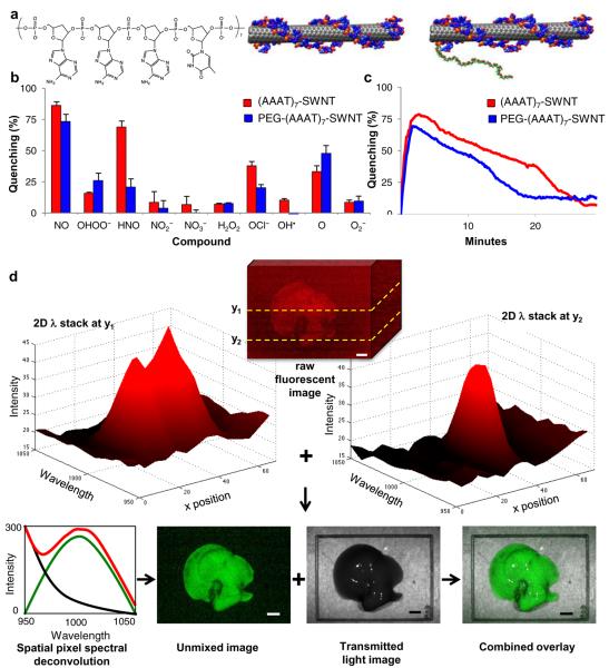
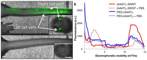
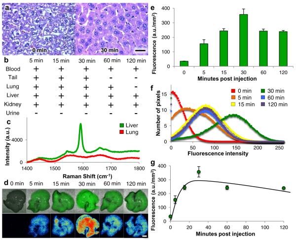
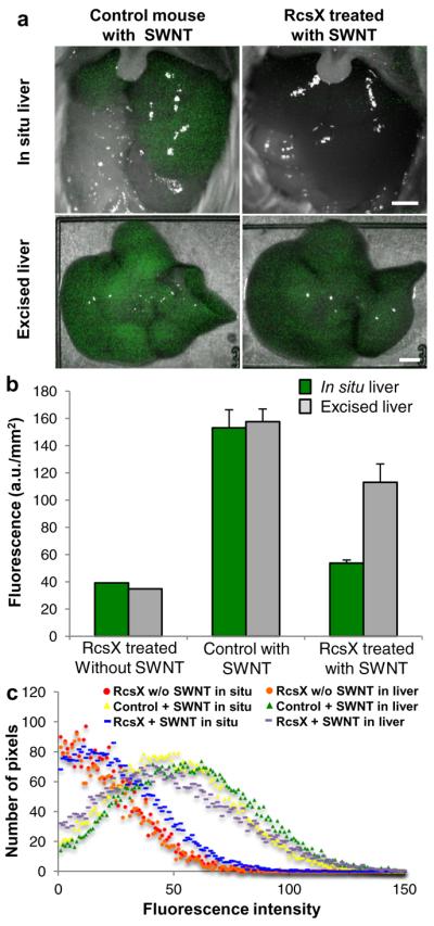
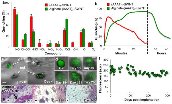
Single-walled carbon nanotubes are particularly attractive for biomedical applications, because they exhibit a fluorescent signal in a spectral region where there is minimal interference from biological media. Although single-walled carbon nanotubes have been used as highly sensitive detectors for various compounds, their use as in vivo biomarkers requires the simultaneous optimization of various parameters, including biocompatibility, molecular recognition, high fluorescence quantum efficiency and signal transduction. Here we show that a polyethylene glycol ligated copolymer stabilizes near-infrared-fluorescent single-walled carbon nanotubes sensors in solution, enabling intravenous injection into mice and the selective detection of local nitric oxide concentration with a detection limit of 1 µM. The half-life for liver retention is 4 h, with sensors clearing the lungs within 2 h after injection, thus avoiding a dominant route of in vivo nanotoxicology. After localization within the liver, it is possible to follow the transient inflammation using nitric oxide as a marker and signalling molecule. To this end, we also report a spatial-spectral imaging algorithm to deconvolute fluorescence intensity and spatial information from measurements. Finally, we demonstrate that alginate-encapsulated single-walled carbon nanotubes can function as implantable inflammation sensors for nitric oxide detection, with no intrinsic immune reactivity or other adverse response for more than 400 days.


Figures

Figure 1
Figure 1. Characterization and 2Dλ imaging analysis of DNA wrapped SWNT complexes
a, chemical composition of complex with d(AAAT)7 and wrapped (AAAT)7-SWNT and PEG-(AAAT)7-SWNT. b,c Quenching activity of (AAAT)7-SWNT (red) and PEG-(AAAT)7-SWNT (blue) sensors quantified by percent quenching of original fluorescence using a 785 nm photodiode following exposure to (b) RNS and ROS compounds (analyzed continuously for 10 minutes and once at the 12 hours post addition time point) with error bars representing standard error (c) NO (analyzed continuously for 30 minutes). (n = 3) d, 2Dλ imaging analysis for (AAAT)7-SWNT in an excised mouse liver 30 minutes after a tail vein injection of PEG-(AAAT)7-SWNT imaged with white light excitation and an emission spectrum from 950 to 1050 nm with a 10 nm step and 20 second accumulation time (scale bars 4 mm).
Figure 2
Figure 2. Effect of PEGylation for tail vein injected SWNT
a, (AAAT)7-SWNT remains within the tail following injection (50 mg L−1) into left then right tail veins, cross-sectional view of tail shows that the (AAAT)7-SWNT is localized within the veins. b, Gel electrophoresis data showing the difference in electrophoretic mobility of (AAAT)7-SWNT (red) when mixed with FBS as opposed to PEG-(AAAT)7-SWNT (blue) which was not altered by the addition of FBS. c, Mouse tail following injection (50 mg L−1) of PEG-(AAAT)7-SWNT into the left tail vein, cross-sectional view shows clearance of SWNT from the vessel. (n = 3, scale bars 2 mm)
Figure 3
Figure 3. Biodistribution and biocompatibility of PEG-(AAAT)7-SWNT in 129 mice
Data from mice sacrificed at various time points following tail vein injection of PEG-(AAAT)7-SWNT (200 μL injection of 50 mg L−1 SWNT) a, Histology images (H&E stained, 60× magnification, scale bar 10 μm) of liver tissue sections for control mouse and mouse received PEG-(AAAT)7-SWNT 30 minutes before sacrifice revealing biocompatibility of SWNT due to lack of inflammatory cell recruitment. b, A representation of SWNT presence (+) or absence (−) in blood, tail (site of injection), lung, liver, kidney and urine following sacrifice and excision (as determined by Raman spectroscopy on fresh samples). c, representative Raman spectrum of those used to determine SWNT localization in (b). d, Images of excised livers deconvoluted with 2Dλ technology showing first PEG-(AAAT)7-SWNT localization relative in the liver then a heatmap of fluorescence (scale bar 4 mm). e, Chart with quantification of SWNT fluorescence in mouse livers excised at various time points following tail vein injection of 200 μL of 50 mg L−1 PEG-(AAAT)7-SWNT. (n = 3–5 mice per time point, error bars are s.e.m.) f, Graph of SWNT fluorescence distribution in mouse livers shown in e, shows similar peak values (10.5, 11.2, 8.1, 9.6 and 10.4) and standard deviations (73.19, 56.43, 63.07, 48.92 and 49.51 a.u.) for all time points. g, Mathematical model of PEG-(AAAT)7-SWNT concentration in mouse liver over time (same livers as those shown in e).
Figure 4
Figure 4. In vivo sensor quenching due to inflammation
a, Inflamed (RcsX treated) and control (healthy) mice imaged in situ 30 minutes after tail vein injection of PEG-(AAAT)7-SWNT (200 μL injection of 50 mg L−1 SWNT) with their livers exposed then the excised liver imaged immediately following sacrifice, displaying the SWNT quenching that is observed in the inflamed animal (scale bars 4 mm). b, Chart showing quantification of SWNT fluorescence, including an extra control of a RcsX treated (inflamed) mouse that received a tail vein injection of saline. (n = 10, error bars are s.e.m.) c, Graph of SWNT fluorescence distribution in mouse livers shown in a and b.
Figure 5
Figure 5. Additional sensor construct with broader in vivo localization possibilities and long term sensing capabilities
a,b Quenching activity of (AAAT)7-SWNT (red) and Alginate-(AAAT)7-SWNT (green) sensors quantified by percent quenching of original fluorescence using a 785 nm photodiode following exposure to (a) RNS and ROS compounds (analyzed continuously for 10 minutes and once at the 12 hours post addition time point) with error bars representing standard error (b) NO (analyzed continuously for 30 minutes for (AAAT)7-SWNT and just under 45 hours for Alginate-(AAAT)7-SWNT) (dashed vertical line represents a change in the timescale from minutes to hours). (n = 3) c, Image of mouse following implantation of two Alginate-(AAAT)7-SWNT gels on day 0 (immediately after implantation of gel 2) and on day 4 after the fluorescence has returned. (scale bar 4 mm) d, Images of Alginate-(AAAT)7-SWNT gel prior to subcutaneous implantation and at various time points throughout the 300 day test period (scale bar 4mm) and (e) quantification of peak SWNT fluorescence for one of the mice tested displaying long term consistency of SWNT signal. f, Histology images (H&E stained, 10x (day 4 and day 400) and 20x (day 180), scale bars 10 μm) from mice sacrificed at three different time points following the subcutaneous implantation of Alginate-(AAAT)7-SWNT show very little to no inflammation.

References

    1. Wray S, Cope M, Delpy DT, Wyatt JS, Reynolds EO. Characterization of the near infrared absorption spectra of cytochrome aa3 and haemoglobin for the non-invasive monitoring of cerebral oxygenation. Biochim Biophys Acta. 1988;933:184–192. - PubMed
    1. Heller DA, et al. Multimodal optical sensing and analyte specificity using single-walled carbon nanotubes. Nat Nanotechnol. 2009;4:114–120. - PubMed
    1. Jin H, Heller DA, Kim JH, Strano MS. Stochastic Analysis of Stepwise Fluorescence Quenching Reactions on Single-Walled Carbon Nanotubes: Single Molecule Sensors. Nano Lett. 2008;8:4299–4304. - PubMed
    1. Jin H, et al. Detection of single-molecule H2O2 signalling from epidermal growth factor receptor using fluorescent single-walled carbon nanotubes. Nat Nanotechnol. 2010;5:302–309. - PMC - PubMed
    1. Kim JH, et al. The rational design of nitric oxide selectivity in single-walled carbon nanotube near-infrared fluorescence sensors for biological detection. Nat Chem. 2009;1:473–481. - PubMed

Publication types