Review
. 2013 Nov 1;5(11):a009076.
doi: 10.1101/cshperspect.a009076.

Biology of the TAM receptors

Affiliations
Review

Biology of the TAM receptors

Greg Lemke. Cold Spring Harb Perspect Biol. .

Abstract

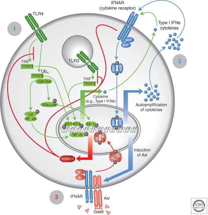
The TAM receptors--Tyro3, Axl, and Mer--comprise a unique family of receptor tyrosine kinases, in that as a group they play no essential role in embryonic development. Instead, they function as homeostatic regulators in adult tissues and organ systems that are subject to continuous challenge and renewal throughout life. Their regulatory roles are prominent in the mature immune, reproductive, hematopoietic, vascular, and nervous systems. The TAMs and their ligands--Gas6 and Protein S--are essential for the efficient phagocytosis of apoptotic cells and membranes in these tissues; and in the immune system, they act as pleiotropic inhibitors of the innate inflammatory response to pathogens. Deficiencies in TAM signaling are thought to contribute to chronic inflammatory and autoimmune disease in humans, and aberrantly elevated TAM signaling is strongly associated with cancer progression, metastasis, and resistance to targeted therapies.


Figures

Figure 1.
Figure 1.
TAM receptors and ligands. The TAM receptors (red) are Tyro3 (Lai and Lemke 1991; Lai et al. 1994)—also designated Brt (Fujimoto and Yamamoto 1994), Dtk (Crosier et al. 1994), Rse (Mark et al. 1994), Sky (Ohashi et al. 1994), and Tif (Dai et al. 1994); Axl (O'Bryan et al. 1991)—also designated Ark (Rescigno et al. 1991), Tyro7 (Lai and Lemke 1991), and Ufo (Janssen et al. 1991); and Mer (Graham et al. 1994)—also designated Eyk (Jia and Hanafusa 1994), Nyk (Ling and Kung 1995), and Tyro12 (Lai and Lemke 1991). The TAMs are widely expressed by cells of the mature immune, nervous, vascular, and reproductive systems. The TAM ligands (blue) are Gas6 and Protein S (Pros1). The carboxy-terminal SHBG domains of the ligands bind to the immunoglobulin (Ig) domains of the receptors, induce dimerization, and activate the TAM tyrosine kinases. When γ-carboxylated in a vitamin-K-dependent reaction, the amino-terminal Gla domains of the dimeric ligands bind to the phospholipid phosphatidylserine expressed on the surface on an apposed apoptotic cell or enveloped virus. See text for details. (From Lemke and Burstyn-Cohen 2010; adapted, with permission, from the authors.)
Figure 2.
Figure 2.
TAM receptor signaling pathways. (A) Free TAMs. As receptor dimers, activated TAM proteins drive a conventional RTK signaling pathway that is dominated by the phosphorylation and activation of Akt. The positions of major tyrosine autophosphorylation sites shared between Tyro3, Axl, and Mer are indicated (P). The tyrosine immediately downstream from the kinase domain (Y821 in human Axl) is bound by the SH2 domain of Grb2, which recruits the p85 subunit of PI3 kinase through an SH3 (Grb2)-proline-rich domain (p85) interaction. Alternatively, p85 can bind this phosphotyrosine directly using its own SH2 domain. P85 also binds to the indicated phosphotyrosine within the kinase domain (see, e.g., Weinger et al. 2008). Mobilization of the joint p85/p110 PI3K complex results in the downstream phosphorylation and activation of Akt. Mer activation has also been found to drive the downstream activation of PLC-γ, by a mechanism that is not delineated biochemically (Tibrewal et al. 2008). These pathways are required for TAM regulation of cell survival and the mobilization of the actin cytoskeleton required for the engulfment of apoptotic cells by phagocytes. (B) TAM receptors complexed with the type I interferon receptor (IFNAR). In dendritic cells, TAM receptors—when activated by the binding of a TAM ligand—form a coimmunoprecipitable complex specifically with the R1 (or α) chain of the IFNAR (Rothlin et al. 2007). This may be associated with the activation of Jak1 (J1) (Zong et al. 1996). Direct activation of the hybrid TAM-IFNAR receptor by the addition of Gas6 leads to the rapid tyrosine phosphorylation and activation of Stat1. This dimeric transcription factor then translocates to the nucleus, where it drives the expression of the cytoplasmic cytokine inhibitors SOCS1 and 3. This pathway is required for the inhibition of inflammatory responses in dendritic cells (Rothlin et al. 2007; Lemke and Rothlin 2008). See text for details.
Figure 3.
Figure 3.
A TAM-regulated cycle of inflammation in dendritic cells. An initial recognition phase (1, green), mediated by Toll-like receptors (e.g., TLR4 on the cell surface and TLR3 in endosomes) and other pattern recognition receptors triggers a kinase cascade that leads to the activation of transcription factors (IRF3/7, AP-1, NF-κB) that drive the production of an initial bolus of type I interferons (IFNs) and other proinflammatory cytokines. In a second response phase (2, blue), the levels of these cytokines are elevated via a feed-forward, JAK-STAT-dependent amplification loop. This same JAK-SAT pathway drives the transcription of the Axl gene. In a final resolution phase (3, red), the induced Axl protein binds to the R1 chain of the type I IFN receptor (IFNAR). The hybrid TAM-IFNAR receptor activates a Stat1 dimer that drives the transcription of the genes encoding SOCS1 and SOCS3. These proteins inhibit both TLR and cytokine receptor signaling, and thereby return the dendritic cell to baseline (Rothlin et al. 2007; Lemke and Rothlin 2008). The features of this self-limiting cycle predict that the provision of an immune stimulus to a dendritic cell with diminished TAM signaling will always result in a hyperelevated inflammatory response. See text for details. (From Rothlin et al. 2007; adapted and reprinted, with permission, from the author.)

References

    1. A-Gonzalez N, Bensinger SJ, Hong C, Beceiro S, Bradley MN, Zelcer N, Deniz J, Ramirez C, Diaz M, Gallardo G, et al. 2009. Apoptotic cells promote their own clearance and immune tolerance through activation of the nuclear receptor LXR. Immunity 31: 245–258 - PMC - PubMed
    1. Ait-Oufella H, Pouresmail V, Simon T, Blanc-Brude O, Kinugawa K, Merval R, Offenstadt G, Leseche G, Cohen PL, Tedgui A, et al. 2008. Defective mer receptor tyrosine kinase signaling in bone marrow cells promotes apoptotic cell accumulation and accelerates atherosclerosis. Arterioscler Thromb Vasc Biol 28: 1429–1431 - PubMed
    1. Akira S 2006. TLR signaling. Curr Top Microbiol Immunol 311: 1–16 - PubMed
    1. Allen MP, Linseman DA, Udo H, Xu M, Schaack JB, Varnum B, Kandel ER, Heidenreich KA, Wierman ME 2002a. Novel mechanism for gonadotropin-releasing hormone neuronal migration involving Gas6/Ark signaling to p38 mitogen-activated protein kinase. Mol Cell Biol 22: 599–613 - PMC - PubMed
    1. Allen MP, Xu M, Linseman DA, Pawlowski JE, Bokoch GM, Heidenreich KA, Wierman ME 2002b. Adhesion-related kinase repression of gonadotropin-releasing hormone gene expression requires Rac activation of the extracellular signal-regulated kinase pathway. J Biol Chem 277: 38133–38140 - PubMed