. 2014 Jan 1;306(1):H142-53.
doi: 10.1152/ajpheart.00438.2013. Epub 2013 Nov 1.

Cardiomyocyte Ogt is essential for postnatal viability

Affiliations

Cardiomyocyte Ogt is essential for postnatal viability

Lewis J Watson et al. Am J Physiol Heart Circ Physiol. .

Abstract

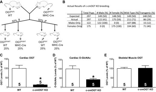
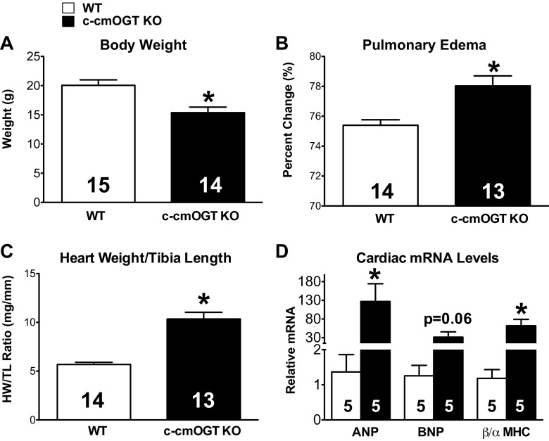
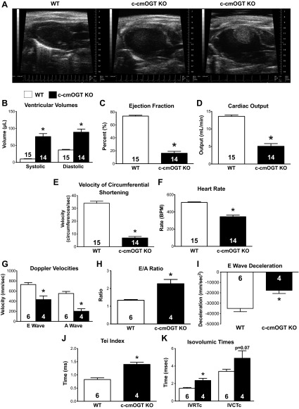
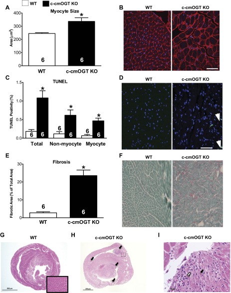
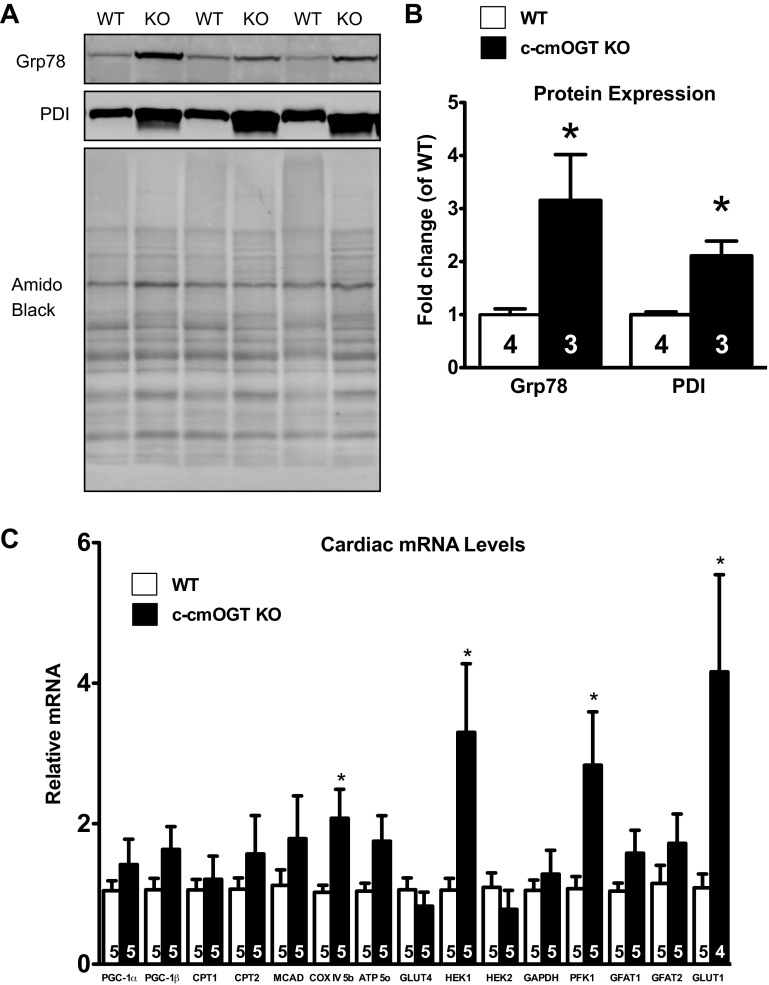
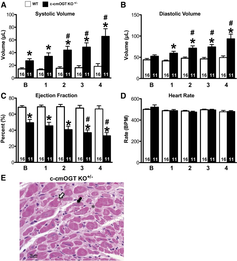
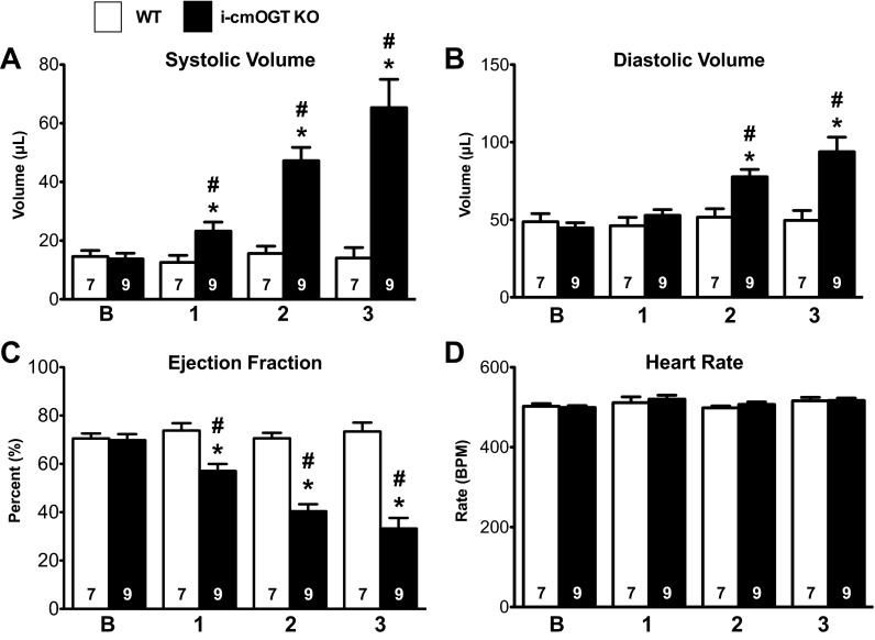
The singly coded gene O-linked-β-N-acetylglucosamine (O-GlcNAc) transferase (Ogt) resides on the X chromosome and is necessary for embryonic stem cell viability during embryogenesis. In mature cells, this enzyme catalyzes the posttranslational modification known as O-GlcNAc to various cellular proteins. Several groups, including our own, have shown that acute increases in protein O-GlcNAcylation are cardioprotective both in vitro and in vivo. Yet, little is known about how OGT affects cardiac function because total body knockout (KO) animals are not viable. Presently, we sought to establish the potential involvement of cardiomyocyte Ogt in cardiac maturation. Initially, we characterized a constitutive cardiomyocyte-specific (cm)OGT KO (c-cmOGT KO) mouse and found that only 12% of the c-cmOGT KO mice survived to weaning age (4 wk old); the surviving animals were smaller than their wild-type littermates, had dilated hearts, and showed overt signs of heart failure. Dysfunctional c-cmOGT KO hearts were more fibrotic, apoptotic, and hypertrophic. Several glycolytic genes were also upregulated; however, there were no gross changes in mitochondrial O2 consumption. Histopathology of the KO hearts indicated the potential involvement of endoplasmic reticulum stress, directing us to evaluate expression of 78-kDa glucose-regulated protein and protein disulfide isomerase, which were elevated. Additional groups of mice were subjected to inducible deletion of cmOGT, which did not produce overt dysfunction within the first couple of weeks of deletion. Yet, long-term loss (via inducible deletion) of cmOGT produced gradual and progressive cardiomyopathy. Thus, cardiomyocyte Ogt is necessary for maturation of the mammalian heart, and inducible deletion of cmOGT in the adult mouse produces progressive ventricular dysfunction.

Keywords: O-linked-β-N-acetylglucosamine transferase; cardiac function; hypertrophy; metabolism; remodeling; uridine diphospho-N-acetylglucosamine:polypeptide β-N-acetylglucosaminyltransferase.


Figures

Fig. 1.
Fig. 1.
Breeding strategy and limited postnatal viability. A: homozygous O-linked-β-N-acetylglucosamine (O-GlcNAc) transferase (Ogt)-floxed (Fl) female mice were crossed with Ogt wild-type (WT) myosin heavy chain (MHC)-Cre transgenic male mice. This cross should have resulted in an even distribution of the MHC transgene (i.e., 50%) across both sexes. All male mice would be hemizygous (effectively homozygous) for the Ogt-floxed allele, whereas all female mice would be heterozygous. B: loss of Ogt did not produce the predicted Mendelian segregation. Tissues were harvested from 4-wk-old animals and subjected to biochemical analysis. C: expression of cardiac OGT was significantly reduced in constitutive cardiomyocyte-specific OGT (c-cmOGT) knockout (KO) mice. D: total cardiac O-GlcNAcylated protein levels were also significantly reduced compared with WT mice. E: to confirm the tissue specificity of Ogt ablation, skeletal muscle was removed and probed for OGT. No change was observed. *P < 0.05 vs. WT mice.
Fig. 2.
Fig. 2.
Gross phenotype of c-cmOGT KO mice. Constitutive ablation of cardiomyocyte Ogt led to gross physical changes. A: c-cmOGT KO mice weighed significantly less than their WT littermates. B: ablation of Ogt led to pulmonary edema. C: c-cmOGT KO mice had enlarged hearts. HW/TL ratio, heart weight-to-tibia length ratio. D: several classic markers of heart failure [atrial natriuretic peptide (ANP) and β-MHC] were also significantly elevated in c-cmOGT KO mice. The brain natriuretic peptide (BNP) elevation trended toward a difference but was P = 0.06. *P < 0.05 vs. WT mice.
Fig. 3.
Fig. 3.
Cardiac ablation of Ogt induces cardiomyopathy. By echocardiography, c-cmOGT KO mice were found to have severe dilated cardiomyopathy (representative echocardiograms are shown in A). B: ablation of cardiac Ogt led to increases in both systolic and diastolic volumes. c-cmOGT KO mice were also found to have decreased ejection fraction (C) and lowered cardiac output (D). E: c-cmOGT KO mice had a slower velocity of shortening, which is a measure of cardiac contractility. F: c-cmOGT KO mice showed a significant reduction in heart rate [in beats/min (BPM)] that was not remedied with anesthetic alterations. G: Doppler survey of the mitral valve revealed that passive (E wave) and active (A wave) filling of the ventricle was impaired. H: ventricles of c-cmOGT KO were filled mostly by passive filling, as denoted by the E-to-A ratio. I: the E wave deceleration time was reduced, suggesting a less compliant ventricle. J: the myocardial performance index (the Tei index) was also increased, suggesting poorer function in c-cmOGT KO mice. K: c-cmOGT KO mice had prolonged isovolumic relaxation and contraction times after correction for heart rate (IVRTc and IVCTc, respectively). *P < 0.05 vs. WT mice.
Fig. 4.
Fig. 4.
Ventricular remodeling in c-cmOGT KO mice. After echocardiography, hearts were removed for histological assessment. c-cmOGT KO mice demonstrated more hypertrophy than their WT counterparts, as determined by wheat germ agglutinin staining (A); representative images are shown in B. Scale bar = 50 μm. C: constitutive ablation of Ogt led to an increase in the rate of TUNEL positivity in the hearts of c-cmOGT KO mice. This increase occurred in both myocyte and nonmyocyte cell fractions; representative images are shown in D. White arrows indicate TUNEL-positive nuclei. Scale bar = 50 μm. c-cmOGT KO mice also had an increase in interstitial fibrosis, as determined by fast green-sirius red staining (E); representative images are shown in F. G: cross-section of the myocardium from a WT (female) mouse showing intact cardiomyocytes (inset). H: multifocal areas of extensive papillary muscle necrosis (arrows) were present within both the right and left ventricular chambers of male c-cmOGT KO mice. I: higher-magnification image of the boxed region from H showing coagulative necrosis of cardiomyocytes (black arrow) and infiltrating neutrophils (white arrow). *P < 0.05 vs. WT mice.
Fig. 5.
Fig. 5.
Cardiomyocyte Ogt deletion produces endoplasmic reticulum (ER) stress and alterations in metabolic gene expression. After echocardiography, hearts were removed and subjected to protein analysis and quantitative RT-PCR for metabolic genes. A: Western blot analysis showing elevated proteins associated with ER stress. Top, 78-kDa glucose-regulated protein (Grp78); middle, protein disulfide isomerase (PDI); bottom, amido black. B: quantification verified the significantly increased levels of Grp78 and PDI in c-cmOGT KO hearts. C: c-cmOGT KO mice had elevated levels of cytochrome-c oxidase (COX), hexokinase (HEK)1, phosphofructokinase (PFK)1, and glucose transporter (GLUT)1. PGC, peroxisome proliferator-activated receptor-γ coactivator; CPT, carnitine palmitoyltransferase; MCAD, medium-chain acyl-CoA dehydrogenase; ATP 5o, ATP synthase, subunit 5; GFAT, glutamine fructose-6-phosphate amidotransferase. *P < 0.05 vs. WT mice.
Fig. 6.
Fig. 6.
Heterozygous female mice exhibit delayed cardiomyopathy and degeneration of cardiomyocytes. Baseline echocardiographic assessment was performed on 2- to 4-mo-old female mice and repeated monthly for 4 mo (x-axis labels).Heterozygous c-cmOGT KO+/− female mice had significantly larger systolic (A) and diastolic (B) volumes. C: ejection fraction was significantly reduced and decreased significantly over time. D: heart rate was not altered and remained stable throughout the 4-wk study. E: myocardial cross-section from a heterozygous female mouse. There were multifocal areas of cardiomyocyte degeneration with nuclear pyknosis (white arrow), cardiomyocyte atrophy (black arrow), and diffuse interstitial fibrosis (*). *P < 0.05 vs. time-matched control mice; #P < 0.05 vs. baseline of the same genotype.
Fig. 7.
Fig. 7.
Cardiomyocyte Ogt is also required for cardiac maintenance. Homozygous Ogt floxed mice with the MerCreMer transgene (i-cmOGT KO mice) were induced with tamoxifen, given a baseline echo after a tamoxifen washout period, and followed via echocardiography monthly for 3 mo. i-cmOGT KO mice were found to have slightly increased systolic (A) and diastolic (B) volumes after 1 mo of Ogt ablation; these increases were persistent and progressive throughout the course of study. C and D: the progressive expansion of ventricular volumes led to a reduction in ejection fraction (C), whereas heart rate remained constant (D). *P < 0.05 vs. WT mice; #P < 0.05 vs. baseline of the same genotype.

References

    1. Agah R, Frenkel PA, French BA, Michael LH, Overbeek PA, Schneider MD. Gene recombination in postmitotic cells. Targeted expression of Cre recombinase provokes cardiac-restricted, site-specific rearrangement in adult ventricular muscle in vivo. J Clin Invest 100: 169–179, 1997 - PMC - PubMed
    1. Belke DD. Swim-exercised mice show a decreased level of protein O-GlcNAcylation and expression of O-GlcNAc transferase in heart. J Appl Physiol 111: 157–162, 2011 - PubMed
    1. Butkinaree C, Park K, Hart GW. O-linked β-N-acetylglucosamine (O-GlcNAc): extensive crosstalk with phosphorylation to regulate signaling and transcription in response to nutrients and stress. Biochim Biophys Acta 1800: 96–106, 2010 - PMC - PubMed
    1. Caldwell SA, Jackson SR, Shahriari KS, Lynch TP, Sethi G, Walker S, Vosseller K, Reginato MJ. Nutrient sensor O-GlcNAc transferase regulates breast cancer tumorigenesis through targeting of the oncogenic transcription factor FoxM1. Oncogene 29: 2831–2842, 2010 - PubMed
    1. Champattanachai V, Marchase RB, Chatham JC. Glucosamine protects neonatal cardiomyocytes from ischemia-reperfusion injury via increased protein-associated O-GlcNAc. Am J Physiol Cell Physiol 291: C178–C187, 2007 - PubMed

Publication types

MeSH terms

Substances

LinkOut - more resources