

Review
doi: 10.1152/physiol.00041.2013.
Emphysema and mechanical stress-induced lung remodeling
Affiliations
- PMID: 24186935
- PMCID: PMC3858211
- DOI: 10.1152/physiol.00041.2013
Item in Clipboard
Review
Emphysema and mechanical stress-induced lung remodeling
Physiology (Bethesda).
2013 Nov.
Abstract
Transpulmonary pressure and the mechanical stresses of breathing modulate many essential cell functions in the lung via mechanotransduction. We review how mechanical factors could influence the pathogenesis of emphysema. Although the progression of emphysema has been linked to mechanical rupture, little is known about how these stresses alter lung remodeling. We present possible new directions and an integrated multiscale view that may prove useful in finding solutions for this disease.
Conflict of interest statement
No conflicts of interest, financial or otherwise, are declared by the author(s).
Figures
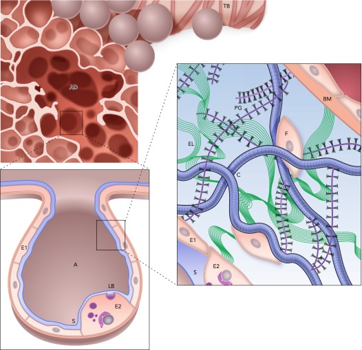
Structure and complexity of the parenchyma at three length scales Top: a terminal bronchiole (TB) leading to an alveolar duct (AD). Bottom left: a zoom into a single air-filled alveolus (A) with type I (E1) and type II (E2) alveolar epithelial cells covered by a thin liquid layer. The surfactant (S) molecules at the air-liquid interface. Secretion of lamellar bodies (LB) by the E2 cell is also shown. Bottom right: a schematic representation of the extracellular matrix of the alveolar septal wall with various components, including amorphous elastin (El), wavy collagen (C), complex proteoglycans (PG), basement membrane (BM), and fibroblast cells (F). Image is adapted from Ref. and is used here with permission from Am J Respir Crit Care Med.
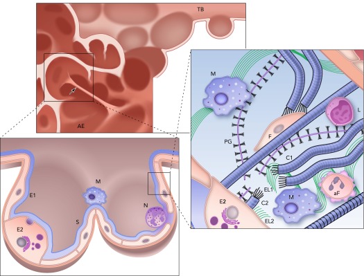
Structure and complexity of the parenchyma at three length scales in the emphysematous lung Top: the remnants of a terminal bronchiole (TB) leading to airspace enlargement (AE). Note the single straight septal wall under larger than average mechanical stress (arrow). Bottom left: zoomed in image of an enlarged airspace region that used to be two alveoli lined with type I (E1) and type II (E2) epithelial cells covered by a thin liquid layer and surfactant (S). Inflammatory cells, including a macrophage (M) and a neutrophil (N), are also shown. Right: a schematic representation of the extracellular matrix of the septal wall with various components including amorphous elastin (El) and collagen (C), and reduced amount of proteoglycans (PG) and fibroblast (F). Compared with the normal ECM in FIGURE 1, here two macrophages (M) and an apoptotic fibroblast (aF) are also seen. Additionally, collagen C is straight because it is under tension, whereas the wavy fragments of collagen C1 and C2 show a rupture site, where elastin (El1 and El2) as well as PG fragments are found. Note also that a macrophage is recruited near this rupture site by the fragments. L denotes a lymphocyte.
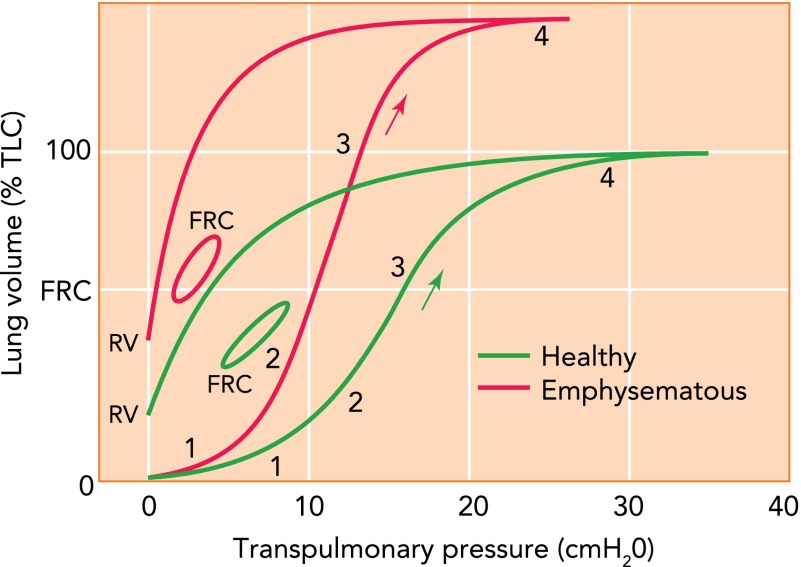
Schematic representation of the pressure-volume curve Schematic representation of the pressure-volume (P-V) curve of a normal (green) and an emphysematous (red) lung during inflation from the collapsed state to total lung capacity (TLC) starting at zero pressure and volume, deflation to residual volume (RV), and during breathing with tidal volume from functional residual capacity (FRC). The regions labeled 1, 2, 3, and 4 correspond approximately to regions of different mechanisms contributing to the curve. The arrow shows the direction of inflation (see text for further explanation). The y-axis is given in percent of TLC of the normal lung.
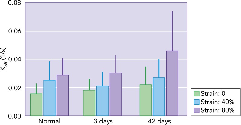
Unbinding rate measured in normal lung tissue and in emphysematous lung tissue Unbinding rate (Koff) measured in normal lung tissue and in emphysematous lung tissue obtained at day 3 or 42 following porcine pancreatic elastase treatment of mice using fluorescent recovery after photobleaching (FRAP), as described in Ref. . Each bar represents the mean and SD of 50–100 individual FRAP curve measurements. The effect of static uniaxial strain on Koff is significant (P < 0.001). Additionally, independent of strain, the effects of treatment are also significant (P < 0.001), with an interaction that almost reached the significance level (P = 0.052) (Sato S, Bartolak-Suki E, Suki B, unpublished observations).
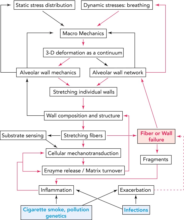
Schematic diagram showing the complexity of multiscale signaling and mechanics with embedded feedback mechanisms influencing ECM composition and lung function in emphysema The arrows represent known or possible links. Notice the feedback from alveolar wall and network mechanics to static stress distribution and dynamic stresses, which decrease transpulmonary pressure and alter breathing pattern. The blue at the bottom represents internal or external triggers. The red pathways show a possible mechanism of self-sustained progression. Fiber and wall failure (red) play a key role here since they directly generate airspace enlargement, feed back to breathing pattern, and, through exposure of fragments, maintain inflammation and drive signaling. The red dashed line represents the added effect of exacerbations on the steady progression.
Comment in
-
Physiology's impact: Applying mathematics and advanced technologies.Physiology (Bethesda). 2013 Nov;28(6):363-5. doi: 10.1152/physiol.00051.2013. Physiology (Bethesda). 2013. PMID: 24186929 No abstract available.
-
Regenerative medicine: why does it matter?Physiology (Bethesda). 2013 Nov;28(6):366-7. doi: 10.1152/physiol.00054.2013. Physiology (Bethesda). 2013. PMID: 24186930 No abstract available.
-
Physiology in perspective: cell migration and the regenerative process.Physiology (Bethesda). 2013 Nov;28(6):368-9. doi: 10.1152/physiol.00052.2013. Physiology (Bethesda). 2013. PMID: 24186931 No abstract available.
References
-
- Alencar AM, Arold SP, Buldyrev SV, Majumdar A, Stamenovic D, Stanley HE, Suki B. Physiology: dynamic instabilities in the inflating lung. Nature 417: 809–811, 2002 - PubMed
-
- Arold SP, Suki B, Alencar AM, Lutchen KR, Ingenito EP. Variable ventilation induces endogenous surfactant release in normal guinea pigs. Am J Physiol Lung Cell Mol Physiol 285: L370–L375, 2003 - PubMed
-
- Baglole CJ, Bushinsky SM, Garcia TM, Kode A, Rahman I, Sime PJ, Phipps RP. Differential induction of apoptosis by cigarette smoke extract in primary human lung fibroblast strains: implications for emphysema. Am J Physiol Lung Cell Mol Physiol 291: L19–L29, 2006 - PubMed
-
- Barnes PJ. Chronic obstructive pulmonary disease. N Engl J Med 343: 269–280, 2000 - PubMed
Publication types
MeSH terms
Grants and funding
LinkOut - more resources
Full Text Sources
Other Literature Sources