. 2014 Feb;28(2):705-14.
doi: 10.1096/fj.13-231274. Epub 2013 Nov 1.

Greatly improved survival and neuroprotection in aquaporin-4-knockout mice following global cerebral ischemia

Affiliations

Greatly improved survival and neuroprotection in aquaporin-4-knockout mice following global cerebral ischemia

Ryuichi Katada et al. FASEB J. 2014 Feb.

Abstract

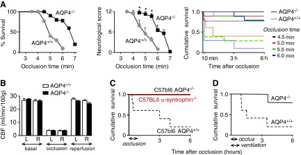
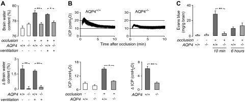
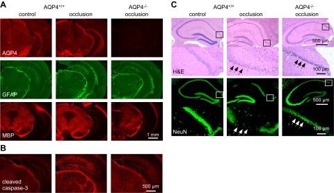
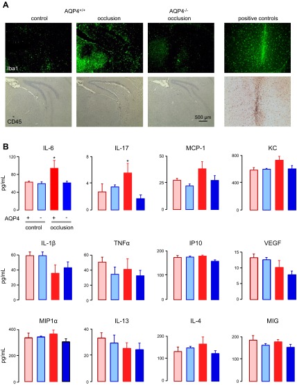
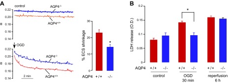
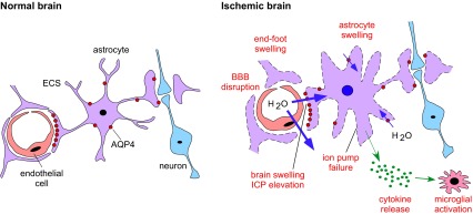
Aquaporin-4 (AQP4), the principal water channel in astrocytes, is involved in brain water movement, inflammation, and neuroexcitation. In this study, there was strong neuroprotection in mice lacking AQP4 in a model of global cerebral ischemia produced by transient, bilateral carotid artery occlusion (BCAO). Survival and neurological outcome were greatly improved in the AQP4(-/-) vs. AQP4(+/+) mice after occlusion, with large and robust differences in both outbred (CD1) and inbred (C57bl/6) mouse strains without or with mechanical ventilation. Improved survival was also seen in mice lacking the scaffold protein α-syntrophin, which manifest reduced astrocyte water permeability secondary to defective AQP4 plasma membrane targeting. Intracranial pressure elevation and brain water accumulation were much reduced in the AQP4(-/-) vs. AQP4(+/+) mice after carotid artery occlusion, as were blood-brain barrier (BBB) disruption and neuronal loss. Brain slices from AQP4(-/-) mice showed significantly reduced cell swelling and cytotoxicity in response to oxygen-glucose deprivation, compared with slices from AQP4(+/+) mice. Our findings suggest that the neuroprotective effect of AQP4 deletion in global cerebral ischemia involves reduced astrocyte swelling and brain water accumulation, resulting in reduced BBB disruption, inflammation, and neuron death. AQP4 water transport inhibition may improve survival and neurological outcome after cardiac arrest and in other conditions associated with global cerebral ischemia.

Keywords: astrocyte; brain swelling; neuroscience; water channel.


Figures

Figure 1.
Figure 1.
Improved survival and neurological outcome in AQP4−/− mice after BCAO. A) AQP4+/+ and AQP4−/− mice on a CD1 genetic background were subjected to BCAO (without mechanical ventilation) for between 3.5 and 7 min. Left panel: percentage survival at 6 h after occlusion: 4.5 min, P = 0.07; 5.0 min, P = 0.011; 5.5 min, P = 0.5; 6.0 min, P = 0.001. A total of 53 AQP4+/+ and 57 AQP4−/− mice were studied. Middle panel: neurological score at 6 h in surviving mice. *P < 0.05. Right panel: cumulative survival as a function of time after occlusion of 4.5, 5.0, 5.5, and 6.0 min. B) CBF in AQP4+/+ and AQP4−/− CD1 mice before and during occlusion and after reperfusion. CBF was measured in the cortex of both the left (L) and right (R) hemispheres (means±se, n=4/group). C) Cumulative survival for AQP4+/+, AQP4−/−, and α-syntrophin−/− mice on a C57bl/6 genetic background after 45 min of BCAO (n=10/group). P = 0.02. D) Survival studies in CD1 mice, as in A, for 30 min of BCAO with mechanical ventilation, during and 15 min after occlusion (n=5/group). P = 0.02.
Figure 2.
Figure 2.
Reduced brain swelling, ICP elevation, and BBB disruption in AQP4−/− mice after BCAO. A) Brain water content was measured in nonventilated (as in Fig. 1A) and ventilated (as in Fig. 1D) CD1 mice at 10 and 15 min after 4.5 and 30 min of BCAO, respectively. Top panel: summary of brain water content for 5 mice/group. Bottom panel: difference in brain water content measured in BCAO vs. control mice. B) ICP was recorded between 0.5 and 10 min after 4.5 min of BCAO in nonventilated CD1 mice. Top panels: representative ICP recordings. Bottom left panel: mean ICP (average over 1–10 min after BCAO; n=4–5/group). Bottom right panel: difference in mean ICP (ΔICP) in BCAO vs. control mice. C) Evans blue extravasation measured in nonventilated CD1 mice at 10 min and 6 h after 4.5 min of occlusion (n=5/group). *P < 0.05, **P < 0.01.
Figure 3.
Figure 3.
Reduced cellular damage in AQP4−/− mice after BCAO. A) AQP4, GFAP, and MBP immunofluorescence in brain sections of nonventilated C57bl/6 mice at 24 h after 30 min of BCAO. Representative of 4 mice/group. B) Immunofluorescence of cleaved caspase-3 (apoptosis marker) in control and in AQP4+/+ and AQP4−/− CD1 mice at 24 h after 5 min of BCAO (representative of 3 mice/group). C) H&E-stained hippocampal sections (top panels) in mice treated as in A, shown together with NeuN immunofluorescence (bottom panels) (representative of 3 mice/group). Higher-magnification images of boxed areas are shown under each image. Arrows indicate loss of NeuN immunofluorescence.
Figure 4.
Figure 4.
Brain inflammatory response after BCAO. A) Iba1 (marker of activated microglia) and CD45 (leukocyte marker) immunostaining of brain sections of nonventilated CD1 mice at 24 h after 5 min of BCAO. Positive controls were brain sections 3 d after intracerebral needle insertion. B) Cytokine concentrations in brain homogenates prepared 6 h after 5 min of BCAO in AQP4+/+ and AQP4−/− CD1 mice (n=5/group). *P < 0.05 vs. AQP4+/+ and AQP4−/− controls.
Figure 5.
Figure 5.
Reduced brain cell swelling and cell death in hippocampal slices from AQP4−/− mice after OGD. A) ECS volume was measured continuously in brain slices by a microfiberoptic fluorescence method. Left panel: representative data for hippocampal slices from AQP4+/+ and AQP4−/− mice without and during OGD. Right panel: percentage of ECS shrinkage, as calculated by relative ECS volume change [1 − (αOGDbasal)] (5 slices from 5 mice/group). *P < 0.05. B) LDH release from organotypic hippocampal slice cultures of AQP4+/+ and AQP4−/− mice after 30 min of OGD and at 6 h after return of slices to normal oxygen–glucose levels (reperfusion; 8 slices/plate insert, 4 inserts/group). *P < 0.05.
Figure 6.
Figure 6.
AQP4-dependent mechanisms of neuroprotection in global cerebral ischemia. In the normal brain, astrocytes occupy a strategic position between capillaries and neurons. Perivascular astrocytic end-feet at the BBB maintain brain ion and water homeostasis. In the ischemic brain, AQP4 facilitates astrocyte swelling and water movement across the BBB, producing multiple deleterious responses (red labels).

References

    1. Fujioka M., Okuchi K., Sakaki T., Hiramatsu K., Miyamoto S., Iwasaki S. (1994) Specific changes in human brain following reperfusion after cardiac arrest. Stroke 25, 2091–2095 - PubMed
    1. Dirnagl U., Iadecola C., Moskowitz M. A. (1999) Pathobiology of ischaemic stroke: an integrated view. Trends Neurosci. 22, 391–397 - PubMed
    1. Moskowitz M. A., Lo E. H., Iadecola C. (2010) The science of stroke: mechanisms in search of treatments. Neuron 67, 181–198 - PMC - PubMed
    1. Papadopoulos M. C., Verkman A. S. (2013) Aquaporin water channels in the nervous system. Nat. Rev. Neurosci. 14, 265–277 - PMC - PubMed
    1. Nielsen S., Nagelhus E. A., Amiry-Moghaddam M., Bourque C., Agre P., Ottersen O. P. (1997) Specialized membrane domains for water transport in glial cells: high-resolution immunogold cytochemistry of aquaporin-4 in rat brain. J. Neurosci. 17, 171–180 - PMC - PubMed

Publication types