. 2014 Jan 17;114(2):266-82.
doi: 10.1161/CIRCRESAHA.113.301720. Epub 2013 Nov 1.

Remodeling of the mononuclear phagocyte network underlies chronic inflammation and disease progression in heart failure: critical importance of the cardiosplenic axis

Affiliations

Remodeling of the mononuclear phagocyte network underlies chronic inflammation and disease progression in heart failure: critical importance of the cardiosplenic axis

Mohamed Ameen Ismahil et al. Circ Res. .

Abstract

Rationale: The role of mononuclear phagocytes in chronic heart failure (HF) is unknown.

Objective: Our aim was to delineate monocyte, macrophage, and dendritic cell trafficking in HF and define the contribution of the spleen to cardiac remodeling.

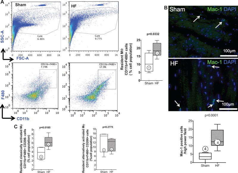
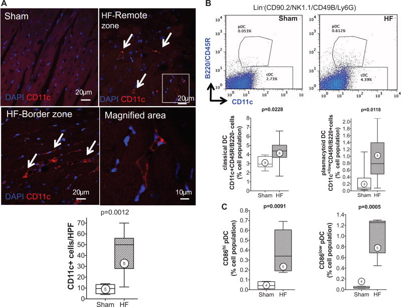
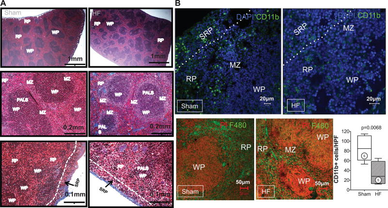
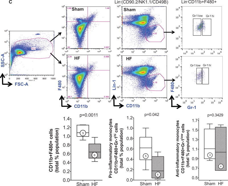
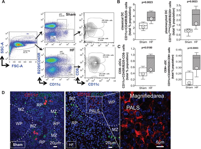
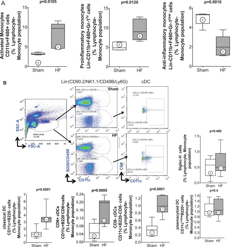
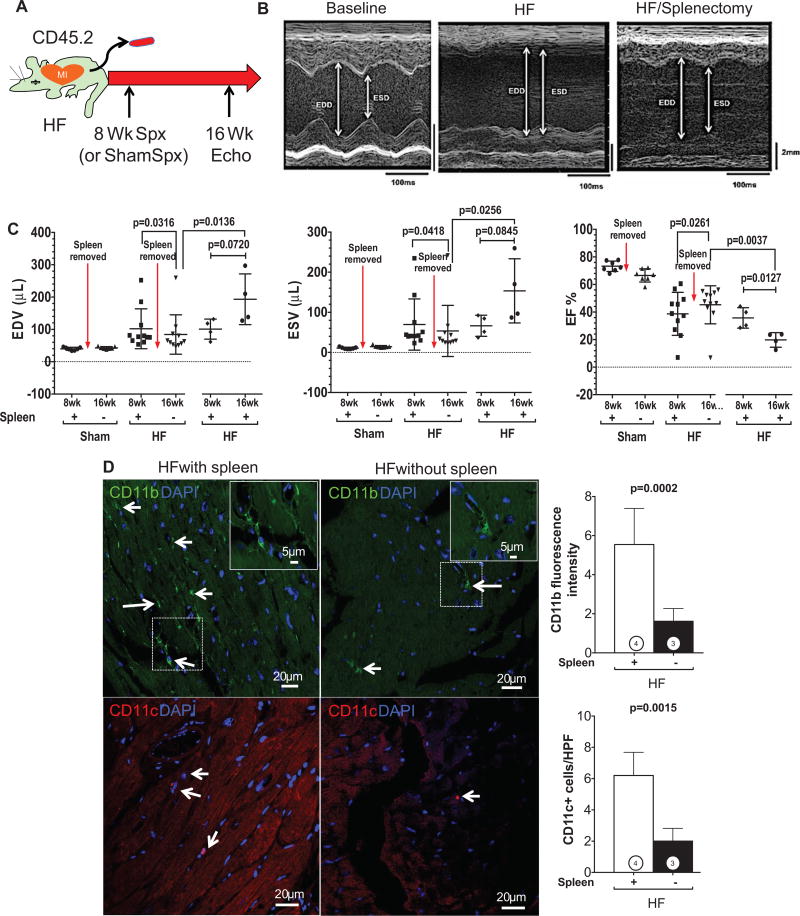
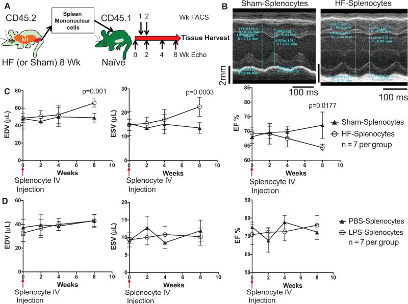
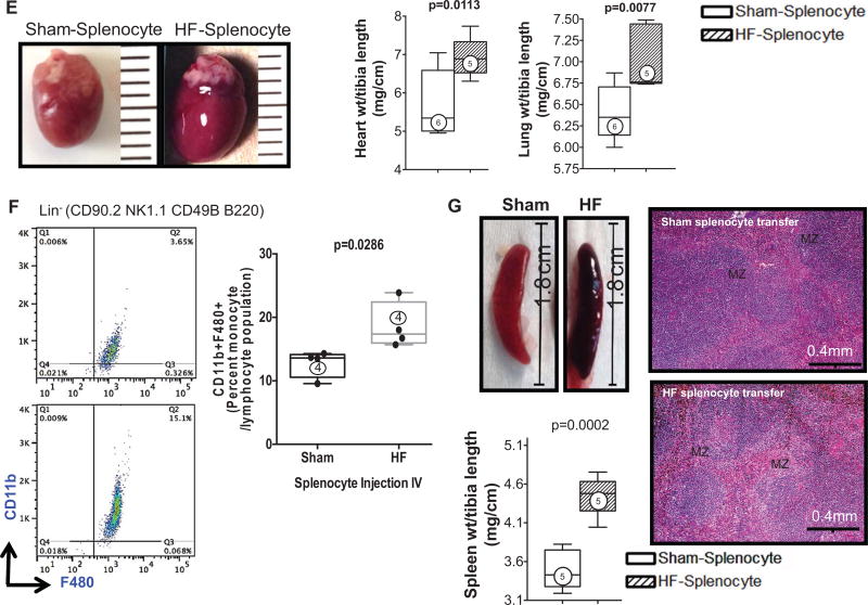
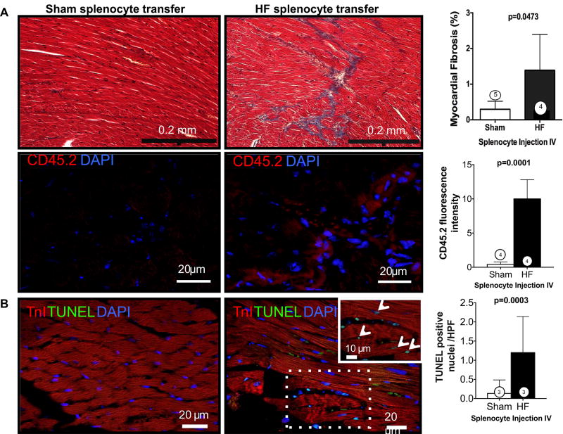
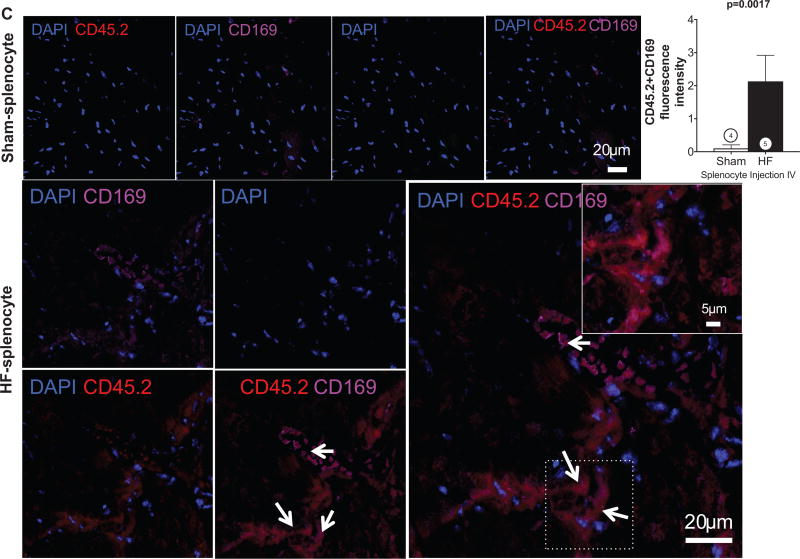
Methods and results: We evaluated C57Bl/6 mice with chronic HF 8 weeks after coronary ligation. As compared with sham-operated controls, HF mice exhibited: (1) increased proinflammatory CD11b+ F4/80+ CD206- macrophages and CD11b+ F4/80+ Gr-1(hi) monocytes in the heart and peripheral blood, respectively, and reduced CD11b+ F4/80+ Gr-1(hi) monocytes in the spleen; (2) significantly increased CD11c+ B220- classical dendritic cells and CD11c+ low)B220+ plasmacytoid dendritic cells in both the heart and spleen, and increased classic dendritic cells and plasmacytoid dendritic cells in peripheral blood and bone marrow, respectively; (3) increased CD4+ helper and CD8+ cytotoxic T-cells in the spleen; and (4) profound splenic remodeling with abundant white pulp follicles, markedly increased size of the marginal zone and germinal centers, and increased expression of alarmins. Splenectomy in mice with established HF reversed pathological cardiac remodeling and inflammation. Splenocytes adoptively transferred from mice with HF, but not from sham-operated mice, homed to the heart and induced long-term left ventricular dilatation, dysfunction, and fibrosis in naive recipients. Recipient mice also exhibited monocyte activation and splenic remodeling similar to HF mice.

Conclusions: Activation of mononuclear phagocytes is central to the progression of cardiac remodeling in HF, and heightened antigen processing in the spleen plays a critical role in this process. Splenocytes (presumably splenic monocytes and dendritic cells) promote immune-mediated injurious responses in the failing heart and retain this memory on adoptive transfer.

Keywords: dendritic cells; heart failure; inflammation; monocytes; spleen.


Figures

Figure 1
Figure 1. Activated macrophages infiltrate the failing heart
A, Cardiac mononuclear cells were isolated on tissue digestion and gradient centrifugation purification. Representative forward and side scatter profiles and live cell gates from a sham and HF heart (top). Identification of Lin2 (defined in text) CD11b+F480+ cells, expressed as a percentage of total live cells, and corresponding group data from sham and HF hearts (bottom). FSC indicates forward scatter; and SSC, side scatter. B, Representative Mac-1 immunostains in sham and HF hearts and corresponding quantification of Mac-1+ cells (arrows). C, Flow cytometric quantification of classically activated M1 (Lin2CD11b+F4/80+CD206) and alternatively activated M2 (Lin2CD11b+F4/80+CD206+) macrophages in sham and HF hearts. Mϕ indicates macrophage; n=4 to 5 per group.
Figure 2
Figure 2. Infiltrating dendritic cells (DCs) are increased in the failing heart
A, Representative confocal images of CD11c immunostained sections from sham and HF hearts (remote zone) and corresponding group data. The arrows indicate infiltrating CD11c+ DCs; n=5 per group. B, Representative scatter plots for Lin1 (defined in text) cardiac mononuclear cells further separated into CD11c+B220 classical DCs (cDCs) and CD11c+/lowB220+ plasmacytoid DCs (pDCs), expressed as a percentage of total live cells, together with quantitative group data for cDC and pDC populations in sham and HF hearts. N=8 per group. C, Flow cytometric quantification of CD86hi and CD86low pDCs in sham and HF hearts. N=4 to 5 per group.
Figure 3
Figure 3. Splenic remodeling in heart failure
A, Representative Masson trichrome stains of spleens from sham-operated and heart failure (HF) mice. The low-power views (top) depict increased white pulp (WP) follicles with greater number of large germinal centers (clear areas in the WP) in the spleen from HF mouse. The higher-power views (middle) illustrate the increased size of the marginal zone (MZ) surrounding the WP in the HF spleen. The bottom panels highlight the subcapsular red pulp (SRP), which exhibits fewer mononuclear cells in HF as compared with sham. PALS indicates periarteriolar lymphoid sheath. B, Representative confocal images of CD11b+ SRP monocytes (top) and F4/80+ monocytes (bottom) in the red pulp of sham and HF spleens, together with quantitative group data for CD11b+ cells in the SRP. C, Representative live cell gates and scatter plots from sham and HF spleens identifying CD11b+F480+ cells, further subdivided by low or high Gr-1 expression (percentage of total live cells), and corresponding group data. N=4 to 6 per group.
Figure 4
Figure 4. Remodeling of the splenic dendritic cell (DC) population in heart failure (HF)
A, Representative scatter plots for splenic mononuclear cells and live cell gates, with subsequent identification of Lin1 CD11c+B220 classical DCs (cDCs) and CD11c+B220+ plasmacytoid DCs (pDCs), and further subdivision of cDCs as CD8+ or CD8 cells. B and C, Quantitative group data for cDCs and pDCs, and CD8+ and CD8 cDCs, expressed as a percentage of total live cell population in sham and HF spleens. N=4 to 6 per group. D, Representative confocal images of CD11c+ DCs in sham and HF spleens demonstrating prominent spatial redistribution of DCs to the white pulp germinal centers in the HF spleen.
Figure 5
Figure 5. Altered circulating mononuclear phagocytes in heart failure (HF)
A, Lin2CD11b+F4/80+ monocytes were identified from the monocyte–lymphocyte gate of peripheral blood leukocytes and further subdivided as high or low Gr-1–expressing cells (pro- and anti-inflammatory monocytes, respectively). Quantitative group data for circulating monocyte subsets in sham and HF mice are shown; n=5 per group. B, Schema for flow cytometric identification of circulating Lin1CD11c+B220 classical DCs (cDCs) and CD11c+B220+ plasmacytoid DCs (pDCs), and CD8+ and CD8 cDCs, expressed as a percentage of the monocyte–lymphocyte gate of peripheral blood cells determined from side and forward scatter (SSC, FSC) plots as shown, together with quantitative group data for the same in sham and HF mice. In parallel studies, peripheral blood pDCs were alternatively identified as Siglec-H+ cells and expressed as a percentage of the monocyte–lymphocyte gate. N=5 to 12 per group.
Figure 6
Figure 6. The spleen influences pathological cardiac remodeling in heart failure (HF)
A, Schema for splenectomy studies in mice with chronic HF. Splenectomy (or sham abdominal surgery) was performed in HF mice 8 weeks after coronary ligation (or sham operation), and remodeling was assessed by echocardiography over an additional 8 weeks. B, M-mode ECGs from 1 mouse at baseline, 8 weeks postligation (HF), and 16 weeks postligation/8 weeks postsplenectomy (HF/splenectomy). C, Quantitative group echocardiographic data for left ventricular (LV) end-diastolic and end-systolic volume (EDV and ESV) and LV ejection fraction (EF) in sham operated and ligated HF mice 8 weeks after splenectomy (n=7 sham mice; n=11 HF mice) or sham abdominal surgery (n=4 HF mice). D. Representative confocal images of CD11b (for macrophages, arrows) and CD11c (for DCs, arrows) immunostained heart sections from HF mice (16 weeks postligation) with or without splenectomy at 8 weeks and corresponding quantification (n=3–4 per group).
Figure 7
Figure 7. Heart failure (HF)-derived splenocytes induce pathological cardiac remodeling on adoptive transfer
A, Schema for splenocyte adoptive transfer experiments. Splenocytes were isolated from CD45.2 sham and HF mice 8 weeks after coronary ligation or sham operation and transferred to syngeneic CD45.1 mice. Recipient mice were then followed for an 8-week period. B, M-mode ECGs from recipient mice 8 weeks after receiving splenocytes from sham or HF mice. C, Serial group echocardiographic data for end-diastolic volume (EDV), end-systolic volume (ESV), and ejection fraction (EF) during the 8-week follow-up period after cell transfer (n=7 per group). D, Similar echocardiographic data in parallel groups of mice after adoptive transfer of splenocytes derived from donors treated with either lipopolysaccharide (LPS) or PBS control (n=7 per group). E, Representative hearts from mice receiving HF or sham splenocytes and corresponding heart and lung gravimetric group data. F, Flow cytometry scatter plots and quantification of circulating CD11b+F4/80+ monocytes in recipient mice 8 weeks after cell transfer. G, Examples of spleens harvested from mice receiving HF or sham splenocytes, corresponding spleen gravimetry, and representative trichrome-stained histological sections from the same. N=4 to 6 per group (E–G).
Figure 8
Figure 8. Trichrome and CD45.2 immunostains (A) and TUNEL stains (B) of hearts from heart failure (HF) or sham splenocyte recipient mice with corresponding quantification of myocardial fibrosis and apoptosis
CD45.2+ donor cells were frequently observed in interstitial and perivascular regions in HF splenocyte recipient hearts (A). C, Representative confocal images of CD45.2- and CD169-immunostained sections from sham and HF splenocyte recipient hearts and corresponding group data for CD45.2+CD169+ cells (arrows). N=3 to 5 per group (A–C).

Comment in

References

    1. Mann DL. Inflammatory mediators and the failing heart: past, present, and the foreseeable future. Circ Res. 2002;91:988–998. - PubMed
    1. Bozkurt B, Mann DL, Deswal A. Biomarkers of inflammation in heart failure. Heart Fail Rev. 2010;15:331–341. - PubMed
    1. Hamid T, Gu Y, Ortines RV, Bhattacharya C, Wang G, Xuan YT, Prabhu SD. Divergent tumor necrosis factor receptor-related remodeling responses in heart failure: role of nuclear factor-kappaB and inflammatory activation. Circulation. 2009;119:1386–1397. - PMC - PubMed
    1. Braunwald E. Biomarkers in heart failure. N Engl J Med. 2008;358:2148–2159. - PubMed
    1. Prabhu SD. Cytokine-induced modulation of cardiac function. Circ Res. 2004;95:1140–1153. - PubMed

Publication types

MeSH terms