. 2013 Nov 1;27(21):2305-19.
doi: 10.1101/gad.228544.113.

Structure and function of Norrin in assembly and activation of a Frizzled 4-Lrp5/6 complex

Affiliations

Structure and function of Norrin in assembly and activation of a Frizzled 4-Lrp5/6 complex

Jiyuan Ke et al. Genes Dev. .

Abstract

Norrin is a cysteine-rich growth factor that is required for angiogenesis in the eye, ear, brain, and female reproductive organs. It functions as an atypical Wnt ligand by specifically binding to the Frizzled 4 (Fz4) receptor. Here we report the crystal structure of Norrin, which reveals a unique dimeric structure with each monomer adopting a conserved cystine knot fold. Functional studies demonstrate that the novel Norrin dimer interface is required for Fz4 activation. Furthermore, we demonstrate that Norrin contains separate binding sites for Fz4 and for the Wnt ligand coreceptor Lrp5 (low-density lipoprotein-related protein 5) or Lrp6. Instead of inducing Fz4 dimerization, Norrin induces the formation of a ternary complex with Fz4 and Lrp5/6 by binding to their respective extracellular domains. These results provide crucial insights into the assembly and activation of the Norrin-Fz4-Lrp5/6 signaling complex.

Keywords: Frizzled 4; Norrin structure; Wnt/β-catenin signaling; cystine knot growth factor; low-density lipoprotein receptor-related protein 5/6; tetraspanin 12.


Figures
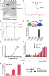
Figure 1.
Figure 1.
MBP-Norrin protein is functional in binding assays and cell-based reporter assays. (A) SDS–gel analysis of purified Fz4-FcH6, Fz8-FcH6, and MBP-Norrin proteins separated under nonreducing (lanes 13) and reducing (lanes 46) conditions. Fz4-FcH6 and Fz8-FcH6 are fusion proteins whose dimerization is mediated through the disulfide bonds in the Fc portion. MBP-Norrin dimerization is mediated through disulfide bonds in the Norrin molecules. (B) Size exclusion column (120 mL of Superdex) profile for MBP-Norrin. MBP-Norrin eluted at 75 mL, corresponding to the size of a dimer. (C) Binding of MBP-Norrin to Fz4-FcH6, Fz8-FcH6, or human IgG was measured by biolayer interferometry. Protein A biosensors were loaded with Fz4-FcH6, Fz8-FcH6, or human IgG. The loaded biosensors were incubated sequentially with buffer in the baseline phase, MBP-Norrin protein in the association phase, and buffer in the dissociation phase. (D) The binding of 10 nM recombinant biotin-MBP-Norrin protein to 20 nM Fz4-FcH6 or Fz8-FcH6 as measured by AlphaScreen assay (n = 3; error bars indicate SD). (E) Saturation binding curve for biotin-MBP-Norrin to 4 nM Fz4-FcH6 as measured by AlphaScreen assay. The Kd was determined as 11 nM by nonlinear regression using GraphPad Prism. (F) Bacterially expressed MBP-Norrin activates TCF-mediated luciferase reporter activity in Fz4-expressing cells. 293STF cells were transfected with the indicated constructs, and luciferase activity was measured as described. (G) Activation of TCF-mediated luciferase activity in cells coexpressing Fz4 and Lrp5 in the presence or absence of MBP-Norrin or Norrin. (H) The hydrophobicity of MBP-Norrin as determined by phase separation assay. MBP-Rhodopsin was used as a control. The MBP-Norrin samples were probed with an anti-Norrin antibody, and MBP-Rhodopsin samples were probed with an anti-MBP antibody.
Figure 2.
Figure 2.
The crystal structure of human Norrin protein. (A) The structure of the Norrin monomer is shown in a rainbow color scheme from the N terminus (blue) to the C terminus (red), with four intramolecular disulfide bonds (C39–C96, C65–C126, C69–C128, and C55–C110) shown as stick models. MBP is omitted for clarity. (B) The structure of a Norrin dimer with the monomers colored green and magenta. (C) The Norrin dimer, showing the three intermolecular disulfide bonds (C93–C95, C95–C93, and C131–C131). (D) The Norrin dimer is further stabilized by intermolecular hydrogen bond interactions between β2′ of one monomer (green) and β2 and β4 from the other monomer (magenta). (E) Hydrophobic F89–I123 interaction at the dimeric interface. (F) Each Norrin dimer binds to two Fz4 CRDs. Fz4-T4L-v5H6 or T4L-v5H6 was immunoprecipitated with v5-agarose beads. Coprecipitated MBP-Norrin and Fz4-FcH6 were detected by tag-specific antibodies.
Figure 3.
Figure 3.
Fz4 receptors dimerize in the absence of Norrin ligand. Shown are the static BRET signals (A) and saturation BRET signals (B) in COS-1 cells expressing both Rlu- and YFP-tagged Fz4 receptors. For A, coexpression of soluble Rlu, soluble YFP, or a structurally unrelated type 2 cholecystokinin receptor (CCK2R) was used as negative controls, and the soluble Rlu-YFP was used as a positive control. The shaded area represents the background signal. (C) The effect of the Norrin ligand on the Fz4 BRET signal by coexpression of wild-type and R41E Norrin constructs and vector control or treatment with MBP-Norrin protein. Plots represent mean ± SEM of data from six independent experiments performed in triplicate. (**) P < 0.01; (#) P < 0.05. (D) Bimolecular complementation experiment showing that Fz4 fused to the nonfluorescent complementary halves of YFP and Fz4-YFP-N and Fz4-YFP-C dimerize and establish a strong YFP fluorescent signal on the cell membrane (arrowheads). Bar, 25 μM. The emission spectrum and anisotropy of this signal were appropriate. (E) Norrin did not induce higher oligomerization of Fz4 receptors detected by three-component BRET assay. The absence of significant BRET from the Rlu-tagged Fz4 coexpressed with Fz4 tagged with YFP-N and YFP-C in the absence or presence of Norrin coexpression. The shaded area represents the background signal as determined using Rlu-tagged Fz4 with CCK2R-YFP. A significant static BRET signal between Rlu- and YFP-tagged Fz4 constructs was used as a positive control. (F) The saturation BRET signals for these complexes, establishing the significance of the signal from the Fz4-Rlu/Fz4-YFP pair and the lack of significance of the signal from coexpression of the Rlu-tagged Fz4 along with Fz4 tagged with YFP-N and YFP-C. Plots represent mean ± SEM of data from four independent experiments performed in triplicate. (**) P < 0.01.
Figure 4.
Figure 4.
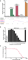
Norrin specifically interacts with Lrp6 BP1–2. (A) AlphaScreen interactions between 30 nM purified biotin-MBP-Norrin and 30 nM indicated proteins. (B) The binding affinity for the interaction between MBP-Norrin and Lrp6 BP12 was determined by a homologous AlphaScreen competition assay using 12 nM Lrp6 BP12 and 120 nM biotinylated MBP-Norrin. The left two bars are negative controls with only single proteins. The last bar shows the signal of the reaction with MBP as a negative competitor control. (C) The competition data were normalized, and the IC50 was determined as 566 nM by nonlinear regression using GraphPad Prism, which corresponds to a Kd of ∼450 nM by Cheng and Prussoff conversion (Cheng and Prusoff 1973).
Figure 5.
Figure 5.
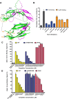
Norrin contains separate binding sites for Fz4 and Lrp5/6. (A) A ribbon diagram of the Norrin dimer with the putative residues involved in binding to Fz4 (blue) or Lrp5/6 (orange) shown as stick models. (B) The effect of mutations of the above surface residues on Norrin function. 293STF cells were transfected with Fz4+Lrp6 in the presence or absence of Norrin wild type (WT) or mutants (n = 3; error bars indicate SD). (C) AlphaScreen competition assay using MBP-Norrin wild-type or mutant proteins to compete the interaction between biotin-MBP-Norrin and Fz4-FcH6 protein. (D) AlphaScreen competition assay using MBP-Norrin wild-type or mutant proteins to compete the interaction between biotinylated DKK1 peptide and H8-Lrp6 BP1–2 protein. IC50 values were derived from curve fitting and are shown in Supplemental Table 1.
Figure 6.
Figure 6.
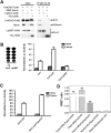
Norrin interacts with Fz4 and Lrp5 to activate downstream β-catenin signaling, and Tspan12 enhances Norrin/Fz4/Lrp5 signaling by interacting with Fz4. (A) Norrin forms a ternary complex with the Fz4 CRD and the Lrp5 ECD. Lrp5NT-v5H6 and T4L-v5H6 (negative control) were immunoprecipitated with v5-agarose beads. Coimmunoprecipitated MBP-Norrin and Fz4-FcH6 were detected by tag-specific antibodies. (B, left) A diagram of full-length Lrp5 and the Lrp5 C-terminal truncation (Lrp5NT). (Right) Lrp5-NT inhibits Norrin-mediated TCF luciferase reporter activity in a dominant-negative manner. 293STF cells were transfected with Fz4, Fz4+Lrp5, or Fz4+Lrp5-NT in the presence or absence of Norrin expression vector. Luciferase activity was measured as described (n = 3; error bars indicate SD). (C) Tspan12 increased Norrin/Fz4/Lrp5-mediated β-catenin signaling. 293STF cells were transfected with Fz4+Lrp5 or Fz4+Lrp5+Tspan12 in the presence or absence of Norrin expression vector. Luciferase activity was measured as described. (n = 3; error bars indicate SD). (D) Tspan12 strongly interacts with Fz4 receptor on the cell membrane. Shown are the static BRET signals in COS-1 cells expressing Rlu-tagged or YFP-tagged Tspan12 and YFP-tagged, YFP-N-tagged, and YFP-C-tagged Fz4 receptors. The shaded area represents the background signal as determined by coexpression of the Rlu-tagged Tspan12 with soluble YFP. Plots represent mean ± SEM of data from three independent experiments performed in triplicate. (**) P < 0.01.
Figure 7.
Figure 7.
A model for Norrin-mediated Fz4 receptor activation. In the absence of Norrin, both Fz4 and Tspan12 form homodimers or oligomers. The interaction of Fz4 with Tspan12 brings Fz4 into tetraspanin-enriched microdomains on the cell membrane. A Norrin dimer binds to an Fz4 dimer with high affinity. Simultaneously, Norrin also binds to Lrp5/6 BP1 and BP2 domains through its two edges (1) or, alternatively, predominantly to the Lrp5/6 BP1 domain (2). In the first binding mode, Norrin binds to one Lrp5/6 molecule, whereas in the second binding mode, Norrin can bind up to two Lrp5/6 molecules. The interaction of Norrin with Fz4 and Lrp5/6 brings their C-terminal tails closer for interaction and activation of the downstream β-catenin signaling pathway.

References

    1. Bailey RL, Herbert JM, Khan K, Heath VL, Bicknell R, Tomlinson MG 2011. The emerging role of tetraspanin microdomains on endothelial cells. Biochem Soc Trans 39: 1667–1673 - PubMed
    1. Berger W 1998. Molecular dissection of Norrie disease. Acta Anat (Basel) 162: 95–100 - PubMed
    1. Bourhis E, Wang W, Tam C, Hwang J, Zhang Y, Spittler D, Huang OW, Gong Y, Estevez A, Zilberleyb I, et al. 2011. Wnt antagonists bind through a short peptide to the first β-propeller domain of LRP5/6. Structure 19: 1433–1442 - PubMed
    1. Bradford MM 1976. A rapid and sensitive method for the quantitation of microgram quantities of protein utilizing the principle of protein-dye binding. Anal Biochem 72: 248–254 - PubMed
    1. Chen ZY, Battinelli EM, Fielder A, Bundey S, Sims K, Breakefield XO, Craig IW 1993. A mutation in the Norrie disease gene (NDP) associated with X-linked familial exudative vitreoretinopathy. Nat Genet 5: 180–183 - PubMed

Publication types

MeSH terms