Review
. 2013 Nov;54(11):1851-4.
doi: 10.2967/jnumed.112.115568.

Acoustic and photoacoustic molecular imaging of cancer

Affiliations
Review

Acoustic and photoacoustic molecular imaging of cancer

Katheryne E Wilson et al. J Nucl Med. 2013 Nov.

Erratum in

  • J Nucl Med. 2014 Jun;55(6):1042

Abstract

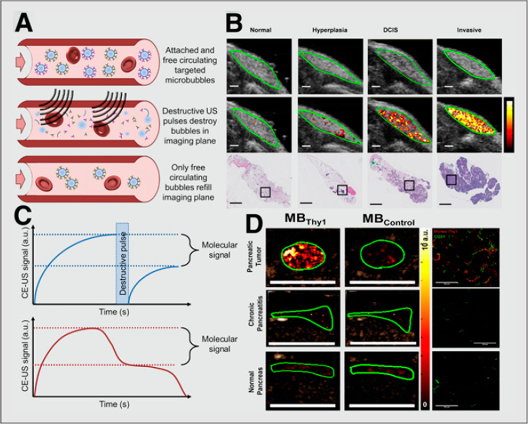
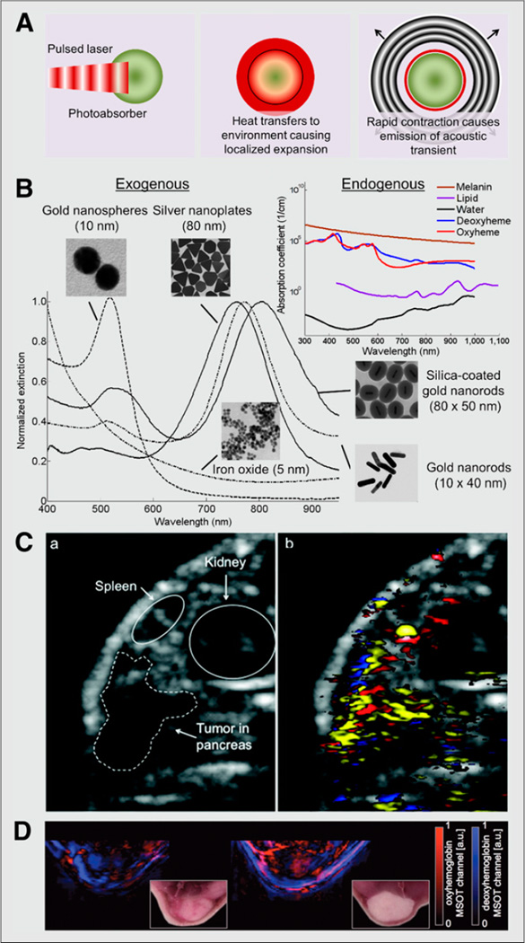
Ultrasound and combined optical and ultrasonic (photoacoustic) molecular imaging have shown great promise in the visualization and monitoring of cancer through imaging of vascular and extravascular molecular targets. Contrast-enhanced ultrasound with molecularly targeted microbubbles can detect early-stage cancer through the visualization of targets expressed on the angiogenic vasculature of tumors. Ultrasonic molecular imaging can be extended to the imaging of extravascular targets through use of nanoscale, phase-change droplets and photoacoustic imaging, which provides further molecular information on cancer given by the chemical composition of tissues and by targeted nanoparticles that can interact with extravascular tissues at the receptor level. A new generation of targeted contrast agents goes beyond merely increasing imaging signal at the site of target expression but shows activatable and differential contrast depending on their interactions with the tumor microenvironment. These innovations may further improve our ability to detect and characterize tumors. In this review, recent developments in acoustic and photoacoustic molecular imaging of cancer are discussed.

Keywords: cancer; microbubble; molecular imaging; nanodroplet; nanoparticle; photoacoustic imaging; ultrasound.


Conflict of interest statement

DISCLOSURE

No potential conflict of interest relevant to this article was reported.

Figures

FIGURE 1
FIGURE 1. Ultrasound molecular imaging of cancer
(A) Schematic showing quantification of ultrasound molecular imaging signal using molecularly targeted contrast microbubbles. (Top) First, microbubbles attach to targets on vascular surface, and signal amplitude is recorded. (Middle) Next, ultrasound pulses are applied to destroy microbubbles within imaging plane. (Bottom) Finally, free circulating microbubbles perfuse into imaging plane and signal amplitude is again recorded. (B) Molecular ultrasound images using clinical-grade human vascular endothelial growth factor receptor 2 (kinase insert domain receptor)–targeted contrast microbubble in transgenic breast cancer mouse model showing increased signal as tissues progress from normal to hyperplasia, ductal carcinoma in situ, and invasive breast cancer (11). (C, top) Difference in pre- and postdestruction images corresponds to signal from attached microbubbles. (C, bottom) Alternative method for determining molecular signal involves waiting (e.g., 10 min after intravenous injection of microbubbles) to allow clearance of freely circulating microbubbles and measuring steady-state signal corresponding to microbubbles attached to molecular targets. (D) Ultrasound molecular image of 4-mm tumor in transgenic pancreatic cancer mouse model using microbubbles targeted at Thy-1 (novel pancreatic cancer target) compared with normal pancreas and chronic pancreatitis tissues (13). a.u. = arbitrary unit; CE-US = contrast-enhanced ultrasound; DCIS = ductal carcinoma in situ; MBThy1 = microbubbles targeted at Thy-1; MBControl = microbubbles targeted at control tissues. (B and D reproduced with permission of (11,13).)
FIGURE 2
FIGURE 2. Photoacoustic molecular imaging of cancer
(A) Diagram of photoacoustic effect. Pulsed laser irradiation is absorbed by photoabsorber, causing localized heating and expansion of directly surrounding environment. During rapid contraction, high-frequency acoustic transient (sound wave) is emitted. (B) Absorption spectra of endogenous and exogenous contrast agents. (C) Ultrasound (left) and spectroscopically resolved photoacoustic images (right) (14.5 × 11.8 mm) of epidermal growth factor receptor targeted silver nanoplates (yellow), oxygenated hemoglobin (red), and deoxygenated hemoglobin (blue) in human pancreatic carcinoma (MPanc96 cells) tumor xenograft (21). (D) Spectroscopically resolved photoacoustic images (and corresponding photographs) of oxy- and deoxyhemoglobin in orthotopic murine breast tumor in mice (20). a.u. = arbitrary unit; MSOT = multispectral optoacoustic tomography. (Silicacoated gold nanorods and iron oxide nanoparticle images and spectra in B reproduced with permission of (24,25); C reproduced with permission of (21); D reproduced with permission of (20).)

References

    1. Kiessling F, Huppert J, Palmowski M. Functional and molecular ultrasound imaging: concepts and contrast agents. Curr Med Chem. 2009;16:627–642. - PubMed
    1. Deshpande N, Needles A, Willmann JK. Molecular ultrasound imaging: current status and future directions. Clin Radiol. 2010;65:567–581. - PMC - PubMed
    1. Unnikrishnan S, Klibanov AL. Microbubbles as ultrasound contrast agents for molecular imaging: preparation and application. AJR. 2012;199:292–299. - PubMed
    1. Dietrich CF, Averkiou MA, Correas J-M, Lassau N, Leen E, Piscaglia F. An EFSUMB introduction into dynamic contrast-enhanced ultrasound (DCE-US) for quantification of tumour perfusion. Ultraschall Med. 2012;33:344–351. - PubMed
    1. Lassau N, Chebil M, Chami L, Bidault S, Girard E, Roche A. Dynamic contrast-enhanced ultrasonography (DCE-US): a new tool for the early evaluation of antiangiogenic treatment. Target Oncol. 2010;5:53–58. - PubMed

Publication types