. 2013 Nov 12;110(46):18472-7.
doi: 10.1073/pnas.1318971110. Epub 2013 Nov 1.

Reconstitution of long and short patch mismatch repair reactions using Saccharomyces cerevisiae proteins

Affiliations

Reconstitution of long and short patch mismatch repair reactions using Saccharomyces cerevisiae proteins

Nikki Bowen et al. Proc Natl Acad Sci U S A. .

Abstract

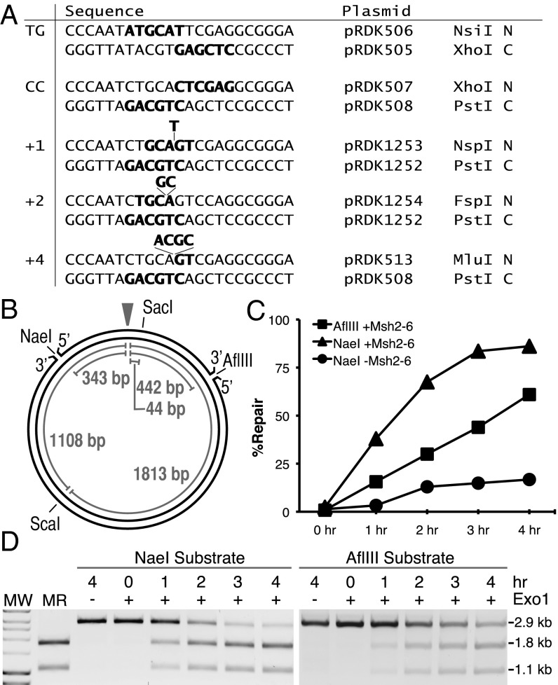
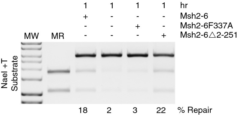
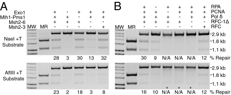
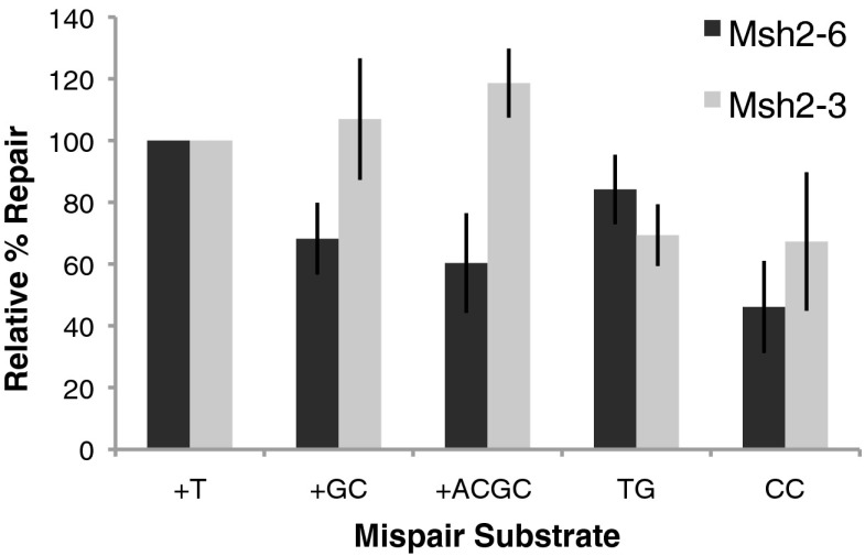
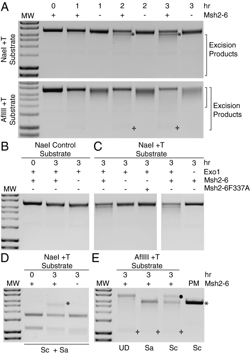
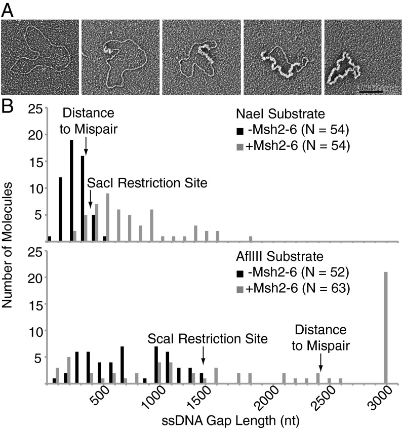
A problem in understanding eukaryotic DNA mismatch repair (MMR) mechanisms is linking insights into MMR mechanisms from genetics and cell-biology studies with those from biochemical studies of MMR proteins and reconstituted MMR reactions. This type of analysis has proven difficult because reconstitution approaches have been most successful for human MMR whereas analysis of MMR in vivo has been most advanced in the yeast Saccharomyces cerevisiae. Here, we describe the reconstitution of MMR reactions using purified S. cerevisiae proteins and mispair-containing DNA substrates. A mixture of MutS homolog 2 (Msh2)-MutS homolog 6, Exonuclease 1, replication protein A, replication factor C-Δ1N, proliferating cell nuclear antigen and DNA polymerase δ was found to repair substrates containing TG, CC, +1 (+T), +2 (+GC), and +4 (+ACGA) mispairs and either a 5' or 3' strand interruption with different efficiencies. The Msh2-MutS homolog 3 mispair recognition protein could substitute for the Msh2-Msh6 mispair recognition protein and showed a different specificity of repair of the different mispairs whereas addition of MutL homolog 1-postmeiotic segregation 1 had no affect on MMR. Repair was catalytic, with as many as 11 substrates repaired per molecule of Exo1. Repair of the substrates containing either a 5' or 3' strand interruption occurred by mispair binding-dependent 5' excision and subsequent resynthesis with excision tracts of up to ~2.9 kb occurring during the repair of the substrate with a 3' strand interruption. The availability of this reconstituted MMR reaction now makes possible detailed biochemical studies of the wealth of mutations identified that affect S. cerevisiae MMR.

Keywords: DNA replication fidelity; cancer; genome instability; mutagenesis; mutator phenotype.


Conflict of interest statement

The authors declare no conflict of interest.

Figures

Fig. 1.
Fig. 1.
Repair of pBluescript-based mispair-containing plasmids in a reconstituted in vitro MMR system. (A) Sequence of the polylinker region between the ApaI and BamHI sites of different substrates indicating the mispair, the restriction sites in each strand, and the plasmid from which each strand was derived. C, continuous strand N, nicked strand. (B) Map of the pBluescript plasmid showing the positions of the various features used in the assays presented and the relevant distances between key sites. The mispair is indicated by the arrowhead. (C and D) Repair of the +1 (+T) substrates containing either a 5′ nick at the NaeI site or a 3′ nick at the AflIII site in reactions for the indicated times containing Msh2–Msh6, Mlh1–Pms1, Exo1, PCNA, RFC-Δ1N, RPA, and DNA polymerase δ with presence/absence of Msh2–Msh6 or Exo1 as indicated. Repair was detected by digestion with PstI and ScaI, and the repair products were visualized after agarose gel electrophoresis (D) and the DNA species seen on the gels were quantified (C). MR, markers for repair products; MW, molecular weight markers. Note: 100% repair is repair of 200 ng or 105.5 fmol of substrate.
Fig. 2.
Fig. 2.
Reconstituted MMR reactions in vitro require the ability of Msh2–Msh6 to bind mispairs but not PCNA. Reconstituted mismatch repair of the +1 (+T) substrate containing a 5′ nick at the NaeI site was performed for 1 h as described in Fig. 1. The presence or absence of Msh2–Msh6, the mispair binding defective Msh2–Msh6–F337A protein, and the PCNA binding defective Msh2–Msh6–Δ2–251 protein and the % repair are as indicated. MR, markers for repair products; MW, molecular weight markers.
Fig. 3.
Fig. 3.
Repair of the +T substrate containing a 5′ nick at the NaeI site or a 3′ nick at the AflIII site in vitro requires Msh2–Msh6 or Msh2–Msh3, and Exo1, PCNA, RFC, RPA, and DNA polymerase δ but not Mlh1–Pms1. Repair of the indicated substrate in 3-h reactions was assayed by digestion with PstI and ScaI as indicated in Fig. 1. The effect of omission of Exo1, Mlh1–Pms1, and Msh2–Msh6 or substitution of Msh2–Msh3 for Msh2–Msh6 is shown in A, and the effect of omission of RPA, PCNA, DNA polymerase δ, and RFC-Δ1N or substitution of RFC for RFC-Δ1N is shown in B. The * in B shows the position of a DNA species formed in the −PCNA, −DNA polymerase δ, and −RFC-Δ1N reactions with the AflIII substrate that has the same mobility as single-stranded pBluescript circular DNA. MR, markers for repair products; MW, molecular weight markers; N/A, no visible repair.
Fig. 4.
Fig. 4.
Mispair specificity of Msh2–Msh6 and Msh2–Msh3 dependent repair. Reactions containing either Msh2–Msh6 or Msh2–Msh3 were performed for 3 h with substrates containing a 5′ nick at the NaeI site and containing +1 (+T), +2 (+GC), +4 (+ACGC), TG, and CC mispairs as indicated (See Figs. 1 and 3). The extent of repair indicated was that relative to the repair of the +1 (+T) substrate present in each set of reactions. The average extent of repair of the +1 (+T) substrate in the Msh2–Msh6 reactions was 49% and in the Msh2–Msh3 reactions was to 67%.
Fig. 5.
Fig. 5.
Excision on the +1 (+T) substrate containing either a 5′ nick at the NaeI site or a 3′ nick at the AflIII site is in the 5′-to-3′ direction and is stimulated by Msh2–Msh6. Excision reactions were performed with +T substrates containing a 5′ nick at the NaeI site or a 3′ nick at the AflIII site in reactions containing Mlh1–Pms1, Exo1, PCNA, RFC-Δ1N, RPA without DNA polymerase δ with or without Msh2–Msh6 or with Msh2–Msh6–F337A as indicated for the indicated times. The reaction products were analyzed by agarose gel electrophoresis without prior digestion with PstI. MW indicates size standards. (A) Excision reactions were performed for 0–3 h. The * indicates a prominent excision product seen with the NaeI substrate in reactions containing Msh2–Msh6. The + shows the position of a DNA species that migrates at the position of single-stranded pBluescript circular DNA seen with the AflIII substrate in reactions containing Msh2–Msh6. (B) Excision reactions performed with a substrate containing a 5′ nick at the NaeI site but lacking a mispair showing the absence of apparent excision products in reactions containing Msh2–Msh6. (C) Excision reactions performed with a +1 (+T) substrate containing a 5′ nick at the NaeI site where Msh2–Msh6 or Exo1 were omitted or the mispair binding defective Msh2–Msh6–F337A protein was substituted for Msh2–Msh6 showing that Msh2–Msh6, mispair binding, and Exo1 were required for the formation of excision products. (D) Analysis of excision products formed with the +T substrate with a 5′ nick at the NaeI site by digestion with SacI (Sa, 387 bp 5′ from the NaeI site) + ScaI (Sc, 2,156 bp 5′ from the NaeI site). The* indicates the full-length linear double-stranded DNA species seen in the presence of Msh2–Msh6. (E) Analysis of excision products formed with the +1 (+T) mispair 3′ nick AflIII substrate by digestion with SacI (Sa, 2,523 bp 5′ from the NaeI site) or ScaI (Sc, 1,371 bp 5′ from the AflIII site) or without digestion (UD). The • indicates the ScaI-resistant nicked circular double-stranded DNA species formed. The * indicates the full-length linear double-stranded DNA species. The + indicates a DNA species that migrates at the position of single-stranded pBluescript circular DNA. PM indicates 100 ng of unincubated substrate DNA digested with ScaI as a control for complete digestion.
Fig. 6.
Fig. 6.
Electron microscopy shows that excision results in single-stranded gaps whose length is stimulated by Msh2–Msh6. Excision reactions were performed for 3 h with +1 (+T) substrates containing either a 5′ nick at the NaeI site or a 3′ nick at the AflIII site in reactions containing Exo1, PCNA, RFC-Δ1N, and RPA without DNA polymerase δ with or without Msh2–Msh6 as indicated. In addition, the reaction with the substrate with the strand interruption at the NaeI site also contained Mlh1–Pms1. (A) A series of representative DNA molecules obtained, with single-stranded DNA stained with E. coli SSB and thus appearing thicker. (Left) Double-stranded circular DNA followed by, from left to right, double-stranded circular DNAs with increasing sizes of single-stranded gaps. (Right) Single-stranded circular DNA. (Scale bar: 100 nm.) (B) Plot of the distribution of single-stranded gap sizes observed. The positions of the mispair, SacI, and ScaI sites are indicated relative to the position of the nick assuming excision is in the 5′ to 3′ direction.

Similar articles

Cited by

References

    1. Hombauer H, Campbell CS, Smith CE, Desai A, Kolodner RD. Visualization of eukaryotic DNA mismatch repair reveals distinct recognition and repair intermediates. Cell. 2011;147(5):1040–1053. - PMC - PubMed
    1. Hombauer H, Srivatsan A, Putnam CD, Kolodner RD. Mismatch repair, but not heteroduplex rejection, is temporally coupled to DNA replication. Science. 2011;334(6063):1713–1716. - PMC - PubMed
    1. Kolodner RD, Marsischky GT. Eukaryotic DNA mismatch repair. Curr Opin Genet Dev. 1999;9(1):89–96. - PubMed
    1. Harfe BD, Jinks-Robertson S. DNA mismatch repair and genetic instability. Annu Rev Genet. 2000;34:359–399. - PubMed
    1. Modrich P, Lahue R. Mismatch repair in replication fidelity, genetic recombination, and cancer biology. Annu Rev Biochem. 1996;65:101–133. - PubMed

Publication types

MeSH terms