

Deferoxamine reduces neuronal death and hematoma lysis after intracerebral hemorrhage in aged rats
- PMID: 24187595
- PMCID: PMC3810989
- DOI: 10.1007/s12975-013-0270-5
Deferoxamine reduces neuronal death and hematoma lysis after intracerebral hemorrhage in aged rats
Abstract
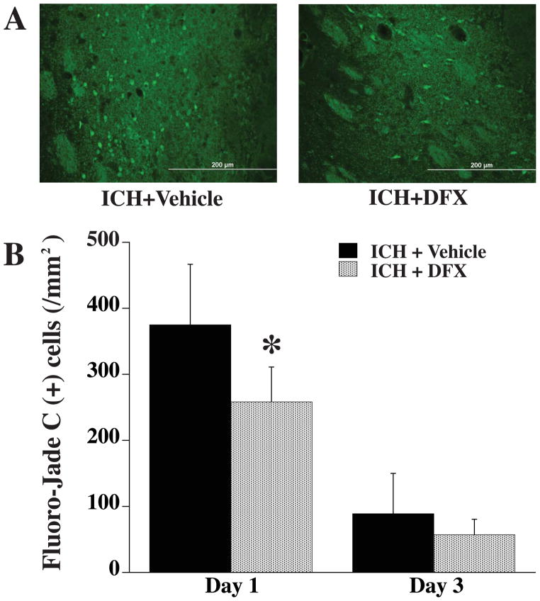
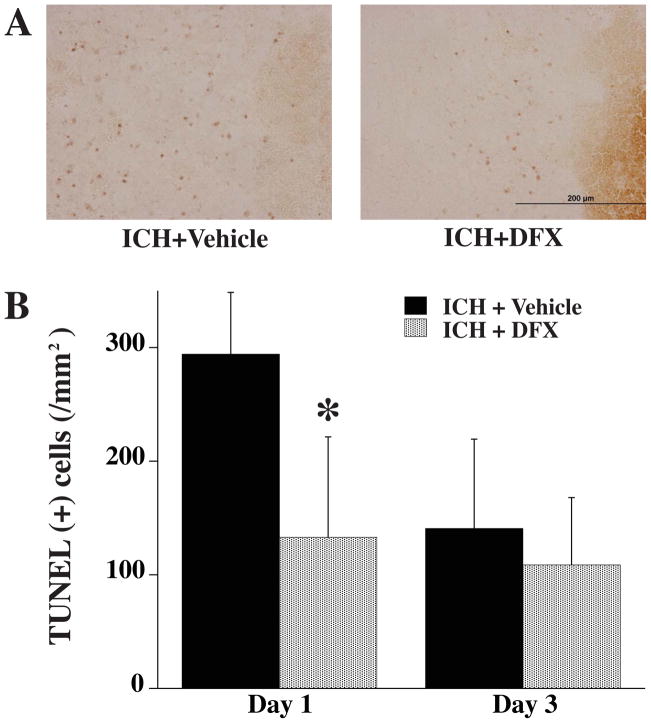
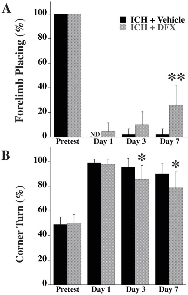
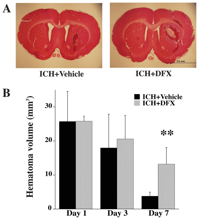
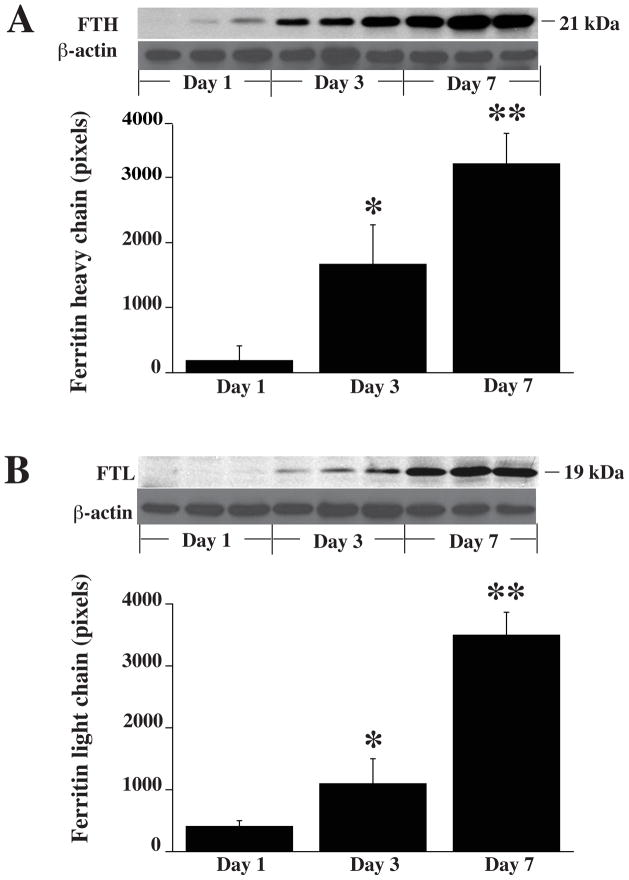
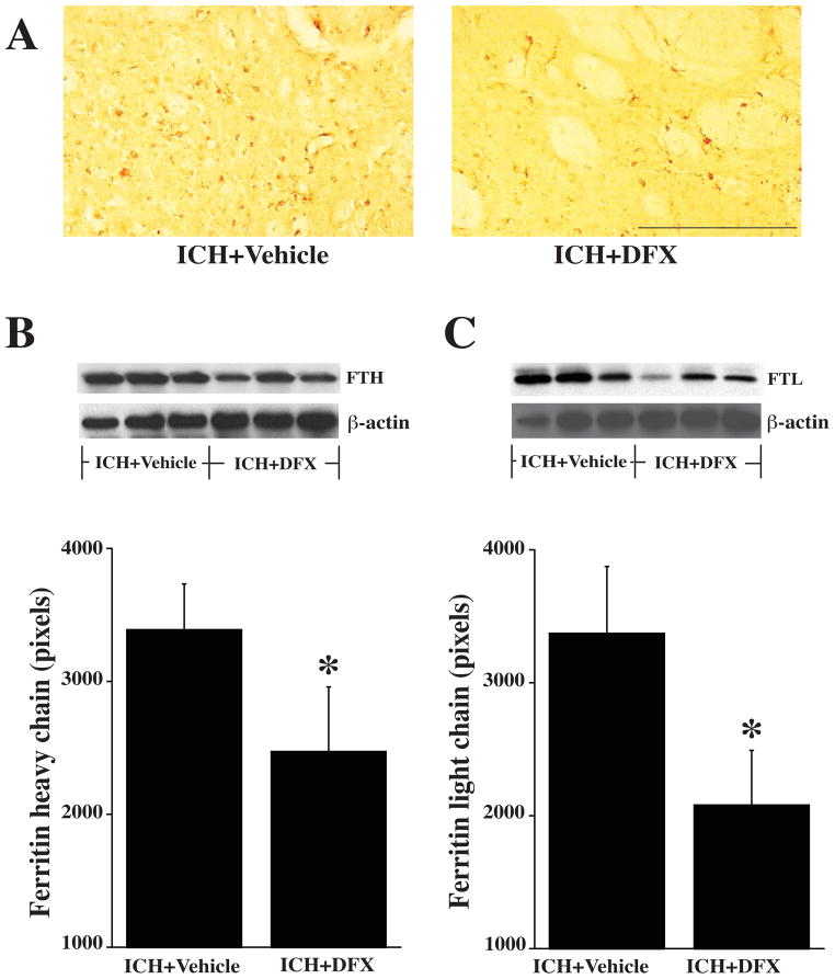
Intracerebral hemorrhage (ICH) is primarily a disease of the elderly. Deferoxamine (DFX), an iron chelator, reduces long-term neurological deficits and brain atrophy after ICH in aged rats. In the present study, we investigated whether DFX can reduce acute ICH-induced neuronal death and whether it affects the endogenous response to ICH (ferritin upregulation and hematoma resolution) in aged rats. Male Fischer 344 rats (18 months old) had an intracaudate injection of 100 μL autologous whole blood into the right basal ganglia and were treated with DFX (100 mg/kg) or vehicle 2 hours post-ICH and then every 12 hours up to 7 days. Rats were euthanized 1, 3, or 7 days later for neuronal death, ferritin and hematoma size measurements. Plasma ferritin levels and behavioral outcome following ICH were also examined. DFX treatment significantly reduced ICH-induced neuronal death and neurological deficits. DFX also suppressed ferritin upregulation in the ipsilateral basal ganglia after ICH and hematoma lysis (hematoma volume at day 7: 13.2±4.9 vs. 3.8±1.2 mm3 in vehicle-treated group, p<0.01). However, effects of DFX on plasma ferritin levels after ICH did not reach significance. In conclusion, DFX reduces neuronal death and neurological deficits after ICH in aged rats. It also affects the endogenous response to ICH.
Keywords: Cerebral hemorrhage; deferoxamine; iron; neuronal death.
Conflict of interest statement
Tetsuhiro Hatakeyama, Masanobu Okauchi, Ya Hua, Richard F. Keep and Guohua Xi declare that they have no conflict of interest.
Figures
References
-
- Huang FP, Xi G, Keep RF, Hua Y, Nemoianu A, Hoff JT. Brain edema after experimental intracerebral hemorrhage: role of hemoglobin degradation products. J Neurosurg. 2002 Feb;96(2):287–93. - PubMed
-
- Wagner KR, Dwyer BE. Hematoma removal, heme, and heme oxygenase following hemorrhagic stroke. Ann N Y Acad Sci. 2004 Mar;1012:237–51. - PubMed
-
- Wagner KR, Sharp FR, Ardizzone TD, Lu A, Clark JF. Heme and iron metabolism: role in cerebral hemorrhage. J Cereb Blood Flow Metab. 2003 Jun;23(6):629–52. - PubMed
-
- Wu J, Hua Y, Keep RF, Schallert T, Hoff JT, Xi G. Oxidative brain injury from extravasated erythrocytes after intracerebral hemorrhage. Brain Res. 2002 Oct 25;953(1–2):45–52. - PubMed
-
- Xi G, Keep RF, Hoff JT. Erythrocytes and delayed brain edema formation following intracerebral hemorrhage in rats. J Neurosurg. 1998 Dec;89(6):991–6. - PubMed
Publication types
MeSH terms
Substances
Grants and funding
LinkOut - more resources
Full Text Sources
Other Literature Sources
Medical
