. 2013 Nov 19;52(46):8187-97.
doi: 10.1021/bi400816s. Epub 2013 Nov 4.

Molecular crowding favors reactivity of a human ribozyme under physiological ionic conditions

Affiliations

Molecular crowding favors reactivity of a human ribozyme under physiological ionic conditions

Christopher A Strulson et al. Biochemistry. .

Abstract

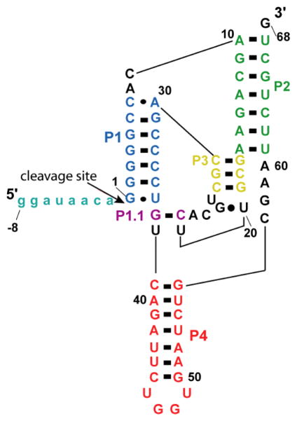
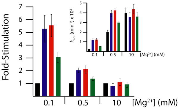
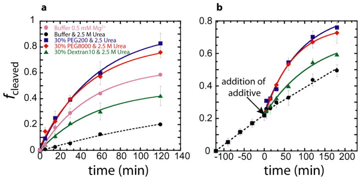
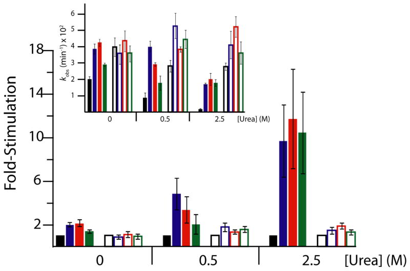
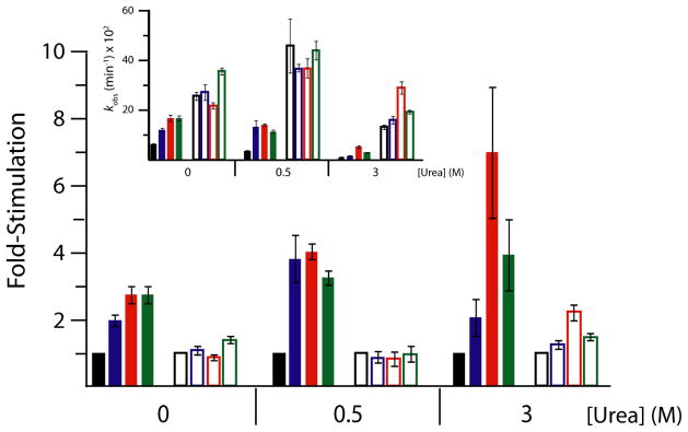
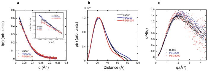
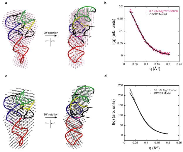
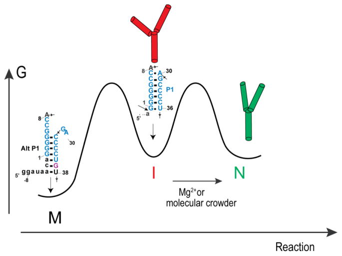
In an effort to relate RNA folding to function under cellular-like conditions, we monitored the self-cleavage reaction of the human hepatitis delta virus-like CPEB3 ribozyme in the background of physiological ionic concentrations and various crowding and cosolute agents. We found that at physiological free Mg(2+) concentrations (∼0.1-0.5 mM), both crowders and cosolutes stimulate the rate of self-cleavage, up to ∼6-fold, but that in 10 mM Mg(2+) (conditions widely used for in vitro ribozyme studies) these same additives have virtually no effect on the self-cleavage rate. We further observe a dependence of the self-cleavage rate on crowder size, wherein the level of rate stimulation is diminished for crowders larger than the size of the unfolded RNA. Monitoring effects of crowding and cosolute agents on rates in biological amounts of urea revealed additive-promoted increases at both low and high Mg(2+) concentrations, with a maximal stimulation of more than 10-fold and a rescue of the rate to its urea-free values. Small-angle X-ray scattering experiments reveal a structural basis for this stimulation in that higher-molecular weight crowding agents favor a more compact form of the ribozyme in 0.5 mM Mg(2+) that is essentially equivalent to the form under standard ribozyme conditions of 10 mM Mg(2+) without a crowder. This finding suggests that at least a portion of the rate enhancement arises from favoring the native RNA tertiary structure. We conclude that cellular-like crowding supports ribozyme reactivity by favoring a compact form of the ribozyme, but only under physiological ionic and cosolute conditions.


Figures

Figure 1
Figure 1. Secondary structure of WT CPEB3 ribozyme
The pairings (P1, P2, P3, P4, and P1.1) of the ribozyme are represented by blue, green, yellow, orange, and purple respectively. Shown is precursor −8/68 WT human RNA, where nucleotides upstream of the cleavage site (−8/−1) are represented in teal.
Figure 2
Figure 2. Stimulation of WT CPEB3 ribozyme kinetics by crowding and cosolute agents in physiological magnesium
Main plot displays relative rate enhancement reported as “fold-stimulation” by comparing rate of condition with additive relative to rate of condition with buffer only (black) for each magnesium concentration, where PEG200 (blue), PEG8000 (red), and Dextran10 (green) were present at a concentration of 30% w/v. Inset: Raw self-cleavage rates of the WT CPEB3 ribozyme for the above-described conditions. Observed cleavage rate constants are provided in Table S1.
Figure 3
Figure 3. Self-cleavage of the WT CPEB3 ribozyme in 0.5 mM Mg2+ and 2.5 M urea with crowding and cosolute agents
PEG200, PEG8000, and Dextan10 were present at final concentrations 30% w/v. (a) Crowding and cosolute additives are present in the reaction solution prior to simultaneous addition of urea and Mg2+. (b) Crowding and cosolute additives were added 120 min after addition of urea and Mg2+, keeping the urea and Mg2+ concentration constant throughout.
Figure 4
Figure 4. Stimulation of WT CPEB3 ribozyme kinetics by crowding and cosolute agents in various concentrations of magnesium and urea
Main plot displays relative rate enhancement reported as “fold-stimulation” by comparing rate of condition with additive relative to rate of condition with buffer only (black) for each Mg2+ concentration, where PEG200 (blue), PEG8000 (red), and Dextran10 (green) are present at concentrations of 30% w/v. Biological free Mg2+ (0.5 mM Mg2+, filled bars) and standard Mg2+ (10 mM Mg2+, open bars) were both used to evaluate ribozyme function in these conditions. Three urea concentrations were chosen to represent varying conditions: 0 M urea (highly folded), 0.5 M urea (denaturing), and 2.5 M urea (highly denaturing). Inset: Raw self-cleavage rates for the WT CPEB3 for the conditions described above. Observed cleavage rate constants are provided in Table S1.
Figure 5
Figure 5. C-2A CPEB3 ribozyme observed cleavage rates with and without crowding and cosolute agents in various concentrations of Mg2+ and urea
Main plot displays relative rate enhancement reported as “fold-stimulation” by comparing rate of condition with additive (colored as below) relative to the condition containing only buffer (black) for each Mg2+ concentration, where PEG200 (blue), PEG8000 (red), and Dextran10 (green) are present at concentrations of 20, 40, and 40%, respectively. Biological free Mg2+ (0.5 mM Mg2+, filled bars) and standard Mg2+ (10 mM Mg2+, open bars) were both used to evaluate the difference between ribozyme function in these conditions. The three urea concentrations were chosen as described in Figure 4. Inset: Raw self-cleavage rates for the C-2A CPEB3 for the conditions described above. Observed cleavage rate values are provided in Table S1.
Figure 6
Figure 6. SAXS scattering profiles, p(r) plots, and Kratky plots in 0.5 mM Mg2+ help describe the global folds
For all panels, plots are for RNA in buffer (black), buffer with 20% PEG200 (blue), and buffer with 20% PEG8000 (red). (a) Scattering profile. RNA scattering profiles in the presence of PEG200 and PEG8000 are normalized to RNA scattering profiles in buffer alone to account for differences in contrast. Rg was determined from data in the Guinier regime (qRg<1.3) (inset), and Rg values are provided in Table 1. Linear behavior extending to low q supports absence of aggregation. (b) p(r) plots. For PEG8000, the distribution is narrower and the peak is at smaller distance which is reflected in a smaller Rg. (c) Dimensionless Kratky plots. These shapes are consistent with a folded native state and absence of significant unstructured regions.
Figure 7
Figure 7. Model of CPEB3 ribozyme agrees well with SAXS data
(a,c) SAXS reconstructions in (a) 0.5 mM Mg2+ and 20% PEG8000 (pink spheres) and (c) 10 mM Mg2+ and no additive (grey spheres) superimposed on the same CPEB3 ribozyme model. The CPEB3 ribozyme model was constructed from the HDV ribozyme crystal structure and color-coded according to the secondary structure in Figure 1. (b,d) Experimental scattering data for CPEB3 ribozyme in (b) 0.5 mM Mg2+ and 20% PEG8000 and (d) 10 mM Mg2+ and no additive plotted with the calculated scattering data from native state model. The calculated scattering profile for CPEB3 model was generated using FoXS server.,
Scheme 1
Scheme 1

References

    1. Strulson CA, Molden RC, Keating CD, Bevilacqua PC. RNA catalysis through compartmentalization. Nat Chem. 2012;4:941. - PubMed
    1. Minton AP. The influence of macromolecular crowding and macromolecular confinement on biochemical reactions in physiological media. J Biol Chem. 2001;276:10577–10580. - PubMed
    1. Ellis RJ. Macromolecular crowding: obvious but underappreciated. Trends Biochem Sci. 2001;26:597–604. - PubMed
    1. Kilburn D, Roh JH, Guo L, Briber RM, Woodson SA. Molecular crowding stabilizes folded RNA structure by the excluded volume effect. J Am Chem Soc. 2010;132:8690–8696. - PMC - PubMed
    1. Nakano S, Karimata HT, Kitagawa Y, Sugimoto N. Facilitation of RNA enzyme activity in the molecular crowding media of cosolutes. J Am Chem Soc. 2009;131:16881–16888. - PubMed

Publication types

LinkOut - more resources