Observational Study
. 2013 Nov 5:13:34.
doi: 10.1186/1471-2342-13-34.

Increased pelvic incidence may lead to arthritis and sagittal orientation of the facet joints at the lower lumbar spine

Affiliations
Observational Study

Increased pelvic incidence may lead to arthritis and sagittal orientation of the facet joints at the lower lumbar spine

Thorsten Jentzsch et al. BMC Med Imaging. .

Abstract

Background: Correct sagittal alignment with a balanced pelvis and spine is crucial in the management of spinal disorders. The pelvic incidence (PI) describes the sagittal pelvic alignment and is position-independent. It has barely been investigated on CT scans. Furthermore, no studies have focused on the association between PI and facet joint (FJ) arthritis and orientation. Therefore, our goal was to clarify the remaining issues about PI in regard to (1) physiologic values, (2) age, (3) gender, (4) lumbar lordosis (LL) and (5) FJ arthritis and orientation using CT scans.

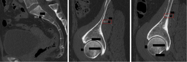
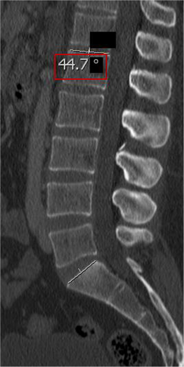
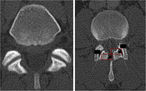
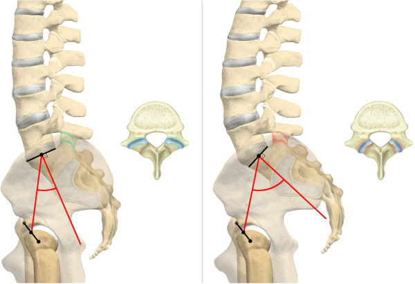
Methods: We retrospectively analyzed CT scans of 620 individuals, with a mean age of 43 years, who presented to our traumatology department and underwent a whole body CT scan, between 2008 and 2010. The PI was determined on sagittal CT planes of the pelvis by measuring the angle between the hip axis to an orthogonal line originating at the center of the superior end plate axis of the first sacral vertebra. We also evaluated LL, FJ arthritis and orientation of the lumbar spine.

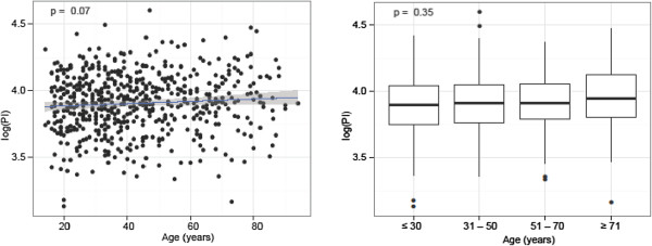
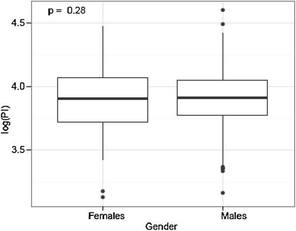
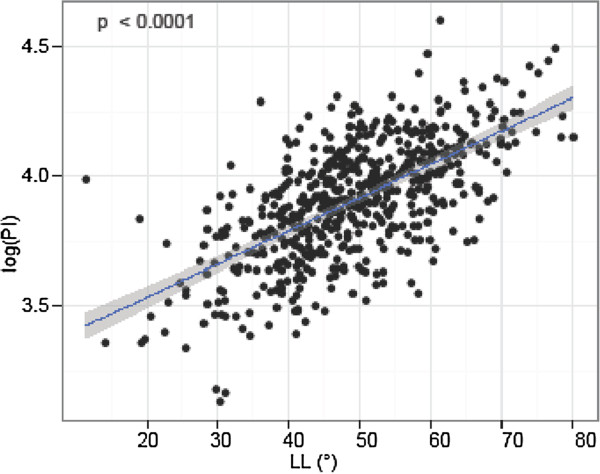
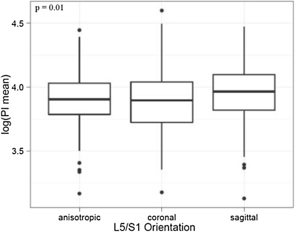
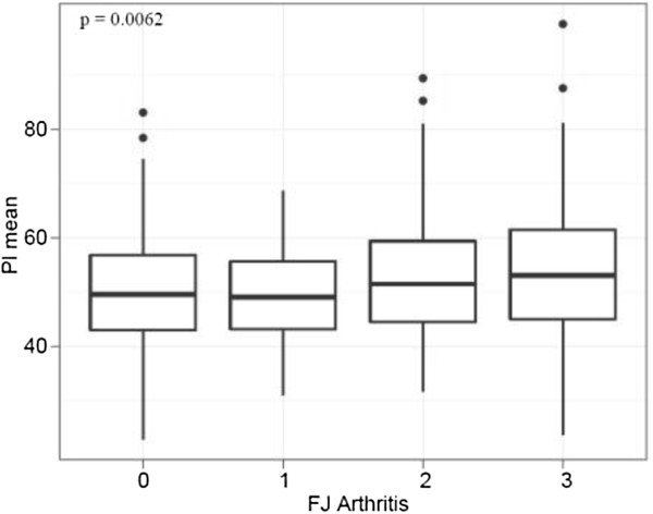
Results: 596 individuals yielded results for (1) PI with a mean of 50.8°. There was no significant difference for PI and (2) age, nor (3) gender. PI was significantly and linearly correlated with (4) LL (p = < 0.0001). Interestingly, PI and (5) FJ arthritis displayed a significant and linear correlation (p = 0.0062) with a cut-off point at 50°. An increased PI was also significantly associated with more sagitally oriented FJs at L5/S1 (p = 0.01).

Conclusion: PI is not correlated with age nor gender. However, this is the first report showing that PI is significantly and linearly associated with LL, FJ arthritis and more sagittal FJ orientation at the lower lumbar spine. This may be caused by a higher contact force on the lower lumbar FJs by an increased PI. Once symptomatic or in the event of spinal trauma, patients with increased PI and LL could benefit from corrective surgery and spondylodesis.


Figures

Figure 1
Figure 1
Pelvic Incidence (PI): The PI was determined on sagittal CT planes of the pelvis. A line was drawn along the axis of the superior end plate of S1 (left image). Then, originating at the center of this axis, an orthogonal line was drawn (left image). Secondly, the middle of the femoral head was determined by the intersecting point of a vertical and horizontal line within the femoral head (middle and right image). Finally, a line was drawn from the middle of the each femoral head to the center of the superior end plate axis and the angle was measured in regard to the orthogonal line originating at this point (middle and right image). The red box indicates the PI. The blacked out numbers were disregarded because they were created automatically by our software and contained irrelevant information. In order to acquire the superposition of the two femoral heads, left and right, the PI was measured for both sides and the mean was stated.
Figure 2
Figure 2
Lumbar Lordosis (LL): LL was evaluated on median sagittal slides by measuring the angle between the superior endplates of L1 and S1, based on the definition of Stokes and the Scoliosis Research Society [[27],[44]]. The red box indicates the PI. The blacked out numbers were disregarded because they were created automatically by our software and contained irrelevant information.
Figure 3
Figure 3
Facet Joints (FJs): FJ orientation was evaluated by measuring the angle between the midline of the sagittal plane and the midline of the FJ as described by Schuller and Mahato [[49],[50]]. Coronal FJ orientation is shown on the left side, whereas sagittal orientation including measurement of FJ orientation is shown on the right side. The red box indicates the PI. The blacked out numbers were disregarded because they were created automatically by our software and contained irrelevant information.
Figure 4
Figure 4
Pelvic Incidence (PI) and Age: There was no significant difference for PI and age.
Figure 5
Figure 5
Pelvic Incidence (PI) and Gender: We did not find a significant difference for PI and gender.
Figure 6
Figure 6
Pelvic Incidence (PI) and Lumbar Lordosis (LL): PI was significantly and linearly correlated with LL.
Figure 7
Figure 7
Pelvic Incidence and FJ Orientation at L5/S1: There was a significant difference in the logarithm of the mean PI and FJ orientation at L5/S1. The FJ orientation was labeled as coronal if angles were > 45° and sagittal if angles were ≤ 45°.
Figure 8
Figure 8
Pelvic Incidence (PI), Facet Joint (FJ) Arthritis and Orientation at L5/S1: On the left side, low PI indicates a normal FJ and more coronal FJ orientation at the lower lumbar spine. Contrarily, the right side shows increased PI with associated FJ arthritis and more sagittal FJ orientation at the lower lumbar spine.
Figure 9
Figure 9
Pelvic Incidence (PI) and Facet Joint (FJ) Arthritis: PI and FJ arthritis displayed a significant linear correlation.

Similar articles

Cited by

References

    1. Le Huec JC, Roussouly P. Sagittal spino-pelvic balance is a crucial analysis for normal and degenerative spine. Eur Spine J. 2011;20(Suppl 5):556–557. - PMC - PubMed
    1. Legaye J, Duval-Beaupère G, Hecquet J, Marty C. Pelvic incidence: a fundamental pelvic parameter for three-dimensional regulation of spinal sagittal curves. Eur Spine J. 1998;7(2):99–103. doi: 10.1007/s005860050038. - DOI - PMC - PubMed
    1. Roussouly P, Gollogly S, Berthonnaud E, Dimnet J. Classification of the normal variation in the sagittal alignment of the human lumbar spine and pelvis in the standing position. Spine. 2005;30(3):346–353. doi: 10.1097/01.brs.0000152379.54463.65. - DOI - PubMed
    1. Vrtovec T, Janssen MM, Pernuš F, Castelein RM, Viergever MA. Analysis of pelvic incidence from 3-dimensional images of a normal population. Spine (Phila Pa 1976) 2012;37(8):479–485. doi: 10.1097/BRS.0b013e31823770af. - DOI - PubMed
    1. Kuntz C, Levin LS, Ondra SL, Shaffrey CI, Morgan CJ. Neutral upright sagittal spinal alignment from the occiput to the pelvis in asymptomatic adults: a review and resynthesis of the literature. J Neurosurg Spine. 2007;6(2):104–112. doi: 10.3171/spi.2007.6.2.104. - DOI - PubMed

Publication types