. 2013 Nov;17(11):1506-12.
doi: 10.1111/jcmm.12149. Epub 2013 Nov 4.

Telocytes in human oesophagus

Affiliations

Telocytes in human oesophagus

Xiaoke Chen et al. J Cell Mol Med. 2013 Nov.

Abstract

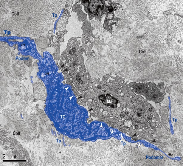
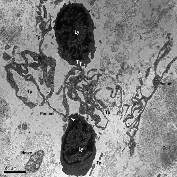
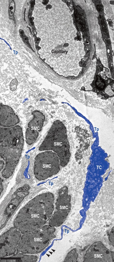
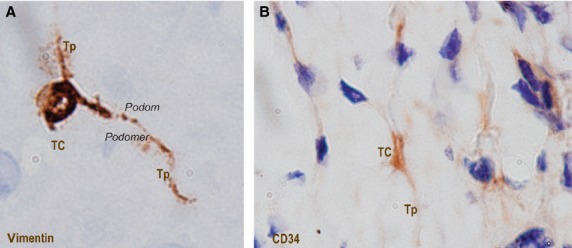
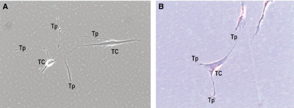
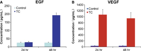
Telocytes (TCs), a new type of interstitial cells, were identified in many different organs and tissues of mammalians and humans. In this study, we show the presence, in human oesophagus, of cells having the typical features of TCs in lamina propria of the mucosa, as well as in muscular layers. We used transmission electron microscopy (TEM), immunohistochemistry (IHC) and primary cell culture. Human oesophageal TCs present a small cell body with 2-3 very long Telopodes (Tps). Tps consist of an alternation of thin segments (podomers) and thick segments (podoms) and have a labyrinthine spatial arrangement. Tps establish close contacts ('stromal synapses') with other neighbouring cells (e.g. lymphocytes, macrophages). The ELISA testing of the supernatant of primary culture of TCs indicated that the concentrations of VEGF and EGF increased progressively. In conclusion, our study shows the existence of typical TCs at the level of oesophagus (mucosa, submucosa and muscular layer) and suggests their possible role in tissue repair.

Keywords: EGF; VEGF; human oesophagus; stromal synapses; telocytes; telopodes; tissue repair.


Figures

Figure 1
Figure 1
Lamina propria of mucosa of human oesophagus. Transmission electron microscopy. Telocyte (TC) and its Telopodes (Tps) in close spatial relationship with a macrophage (Ma). TC extends its Tps wrapping Ma. The typical silhouette of Tps is obvious: alternation of podoms (dilated segments) and podomers (thin segments). Several direct membrane-to-membrane contacts are visible, either point contacts (black arrow-heads), or planar contacts (white arrow-heads). Coll: collagen; m: mitochondria.
Figure 2
Figure 2
Lamina propria of mucosa of human oesophagus. Transmission electron microscopy. Telopodes (Tp), probably of different Telocytes, having a labyrinthine spatial arrangement. The characteristic alternation of podoms and podomers is obvious. Several point contacts (white arrows) are present between Tps and lymphocytes (Ly), at different levels. Even if not in direct contact with nerve, its close vicinity should not be neglected. coll: collagen.
Figure 3
Figure 3
Muscular layer of human oesophagus. Transmission electron microscopy. Telocyte (TC) with two visible Telopodes (Tps) in close vicinity of smooth muscle cells (SMC), and wrapping them. Fragments of (most probably) the same Tp are interposed between SMC and a blood capillary. Small electron-dense nanostructures (arrow-heads) are seen between both membranes – SMC and Tp.
Figure 4
Figure 4
Submucosa of human oesophagus. Immunohistochemistry. Vimentin-positive cells (A) and CD34-positive cells (B). Both cells morphologies are very evocative for Telocytes (TC): small cell body (A and B) with long cell processes (A and B) – Telopodes (Tp), with their characteristic moniliform silhouette (alternation of podoms and podomers) are clearly visible (A). Original magnification: 1000×.
Figure 5
Figure 5
Primary culture of human oesophagus Telocytes. Phase-contrast microscopy. Vimentin-positive cells (A) and CD34-positive cells (B). Both cell morphologies are very evocative for Telocytes (TC): small cell body (A and B) with long cell processes (A and B) – Telopodes (Tp), with their characteristic moniliform silhouette (alternation of podoms and podomers), are visible (A). Original magnification: 1000×.
Figure 6
Figure 6
The concentrations of EGF and VEGF detected by ELISA. (A) The concentration of EGF in the supernatant of cultured telocytes was slightly changed at 24 hrs, but abruptly increased (almost four times) at 48 hrs, in comparison with control. (B) The concentration of VEGF in the supernatant of cultured telocytes was significantly higher than in control at 24 hrs, and slightly decreased at 48 hrs.

References

    1. Popescu LM, Faussone-Pellegrini MS. TELOCYTES - a case of serendipity: the winding way from Interstitial Cells of Cajal (ICC), via Interstitial Cajal-Like Cells (ICLC) to TELOCYTES. J Cell Mol Med. 2010;14:729–40. - PMC - PubMed
    1. Suciu L, Popescu LM, Gherghiceanu M, et al. Telocytes in human term placenta: morphology and phenotype. Cells Tissues Organs. 2010;192:325–39. - PubMed
    1. Gherghiceanu M, Popescu LM. Cardiomyocyte precursors and telocytes in epicardial stem cell niche: electron microscope images. J Cell Mol Med. 2010;14:871–7. - PMC - PubMed
    1. Popescu LM, Manole CG, Gherghiceanu M, et al. Telocytes in human epicardium. J Cell Mol Med. 2010;14:2085–93. - PMC - PubMed
    1. Gherghiceanu M, Manole CG, Popescu LM. Telocytes in endocardium: electron microscope evidence. J Cell Mol Med. 2010;14:2330–4. - PMC - PubMed

Publication types

MeSH terms

Substances

LinkOut - more resources