. 2013 Dec 13;288(50):36160-7.
doi: 10.1074/jbc.M113.524355. Epub 2013 Nov 4.

A highly conserved cysteine of neuronal calcium-sensing proteins controls cooperative binding of Ca2+ to recoverin

Affiliations

A highly conserved cysteine of neuronal calcium-sensing proteins controls cooperative binding of Ca2+ to recoverin

Matthew J Ranaghan et al. J Biol Chem. .

Abstract

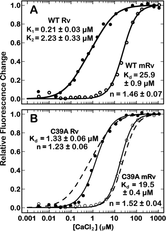
Recoverin, a 23-kDa Ca(2+)-binding protein of the neuronal calcium sensing (NCS) family, inhibits rhodopsin kinase, a Ser/Thr kinase responsible for termination of photoactivated rhodopsin in rod photoreceptor cells. Recoverin has two functional EF hands and a myristoylated N terminus. The myristoyl chain imparts cooperativity to the Ca(2+)-binding sites through an allosteric mechanism involving a conformational equilibrium between R and T states of the protein. Ca(2+) binds preferentially to the R state; the myristoyl chain binds preferentially to the T state. In the absence of myristoylation, the R state predominates, and consequently, binding of Ca(2+) to the non-myristoylated protein is not cooperative. We show here that a mutation, C39A, of a highly conserved Cys residue among NCS proteins, increases the apparent cooperativity for binding of Ca(2+) to non-myristoylated recoverin. The binding data can be explained by an effect on the T/R equilibrium to favor the T state without affecting the intrinsic binding constants for the two Ca(2+) sites.

Keywords: Allosteric Regulation; Calcium Signaling; Calcium-binding Proteins; Cooperativity; Cysteine; Mutant; Neuronal Calcium Sensor; Protein Myristoylation; Sulfenic Acid.


Figures

FIGURE 1.
FIGURE 1.
A, schematic model of mRv (PDB ID code 1IKU) showing the arrangement of color-coded EF hands, the location of Cys-39 in EF-hand 1 (purple), and the N-terminally attached myristoyl group (orange, space filling). B, sequence alignment of the four EF hand loop regions of recoverin. The consensus sequence for EF hand loops is shown with the Ca2+-coordinating residues denoted as X, Y, and Z (underline indicates the negative position), n represents any non-polar residue, and a dash is any residue. The conserved Ca2+-binding residues are marked in red boldface. The gray box in EF1 highlights the conserved CPXG motif.
SCHEME 1
SCHEME 1
FIGURE 2.
FIGURE 2.
Normalized change in fluorescence for Ca2+ binding to myristoylated (open) and non-myristoylated (filled) WT (A) or C39A (B) recoverin. Equation 1 was used to fit titration data for WT mRv, C39A mRv, and C39A Rv. Equation 2 was used to fit the titration data for WT Rv. The dashed lines in B represent the WT titration data from A for comparison.
FIGURE 3.
FIGURE 3.
Binding assays for WT or C39A Rv to RGS. A, pulldown assay using a Ni-NTA matrix to immobilize RGS as described under “Experimental Procedures.” The RGS-Rv complex was eluted with 250 mm imidazole. Lanes are defined as L, load; FT, flow-through; W, last wash; E, imidazole elution. B, ITC isotherm showing the heat observed upon injecting aliquots of WT Rv (660 μm) into a solution of RGS (60 μm) at 30 °C. C, ITC isotherm showing the heat observed upon injecting aliquots of C39A (340 μm) into a solution of RGS (30 μm) at 30 °C.
FIGURE 4.
FIGURE 4.
A and B, normalized change in fluorescence for Ca2+-binding titrations for P40A Rv (A) and C39D Rv (B). Equation 2 was used to fit the titration data. The dashed lines are for WT Rv (from Fig. 2A). C, pulldown assay in which RGS was first immobilized on a Ni-NTA matrix, WT, C39D, or P40A Rv added in the presence of 1 mm CaCl2, and finally the RGS-Rv complex eluted with 250 mm imidazole. Lanes are defined as L, load; FT, flow-through; W, last wash; E, imidazole elution.
FIGURE 5.
FIGURE 5.
A, superposition of the C39A Rv (magenta) structure with that of WT Rv (green). Ca2+ ions are shown as spheres occupying EF3. B, superposition of EF2 (left) and EF3 (right) for C39A and WT Rv. Colors are the same as in A. The red sphere is an axial water molecule.
FIGURE 6.
FIGURE 6.
Electron density at position 39 for WT (A), oxWT (B), C39A (C), and C39D Rv (D). The Fourier map (cyan) is shown at 1 σ cutoff for all the atoms. The omit map (red) is shown at 3 σ cutoff for the O atom of sulfenic acid (B) and the side chain of Asp-39 (D).

Similar articles

Cited by

References

    1. Chen C. K., Woodruff M. L., Chen F. S., Chen D., Fain G. L. (2010) Background light produces a recoverin-dependent modulation of activated-rhodopsin lifetime in mouse rods. J. Neurosci. 30, 1213–1220 - PMC - PubMed
    1. Chen C. K., Woodruff M. L., Chen F. S., Chen Y., Cilluffo M. C., Tranchina D., Fain G. L. (2012) Modulation of mouse rod response decay by rhodopsin kinase and recoverin. J. Neurosci. 32, 15998–16006 - PMC - PubMed
    1. Klenchin V. A., Calvert P. D., Bownds M. D. (1995) Inhibition of rhodopsin kinase by recoverin: further evidence for a negative feedback system in phototransduction. J. Biol. Chem. 270, 16147–16152 - PubMed
    1. Chen C. K., Inglese J., Lefkowitz R. J., Hurley J. B. (1995) Ca2+-dependent interaction of recoverin with rhodopsin kinase. J. Biol. Chem. 270, 18060–18066 - PubMed
    1. Higgins M. K., Oprian D. D., Schertler G. F. (2006) Recoverin binds exclusively to an amphipathic peptide at the N terminus of rhodopsin kinase, inhibiting rhodopsin phosphorylation without affecting catalytic activity of the kinase. J. Biol. Chem. 281, 19426–19432 - PubMed

Publication types

LinkOut - more resources