. 2013 Dec 13;288(50):36029-39.
doi: 10.1074/jbc.M113.525618. Epub 2013 Nov 4.

The wing of a winged helix-turn-helix transcription factor organizes the active site of BirA, a bifunctional repressor/ligase

Affiliations

The wing of a winged helix-turn-helix transcription factor organizes the active site of BirA, a bifunctional repressor/ligase

Vandana Chakravartty et al. J Biol Chem. .

Abstract

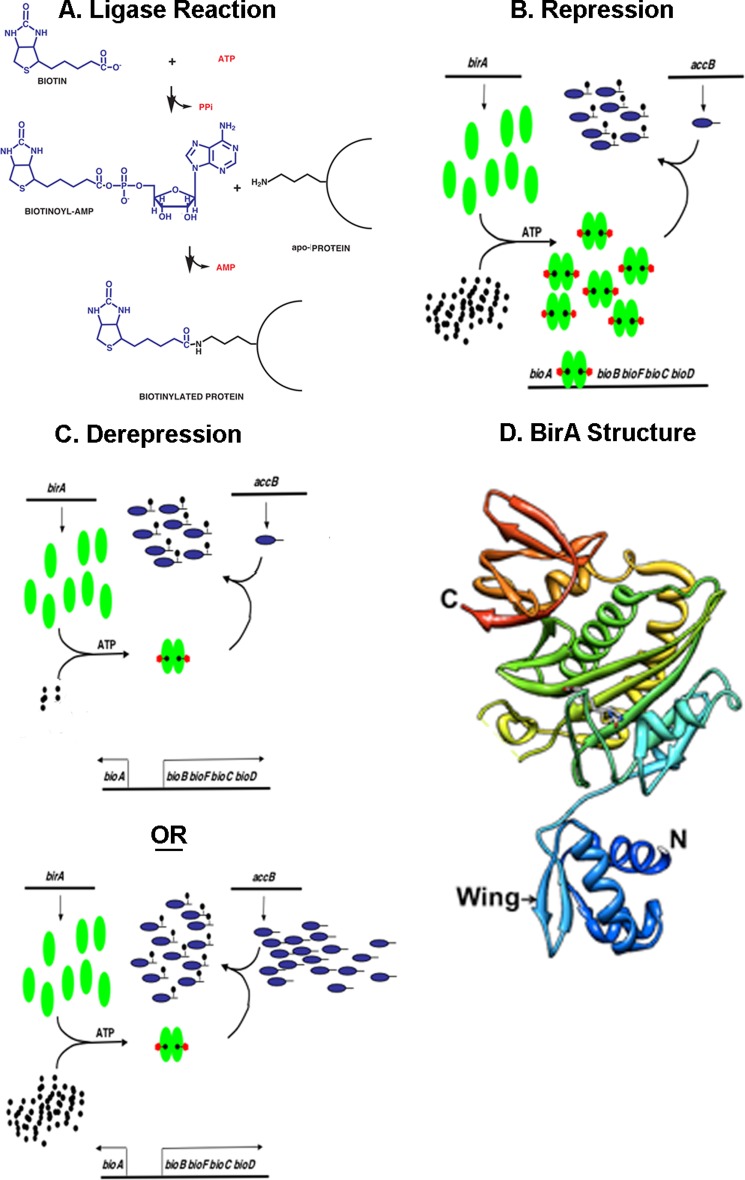
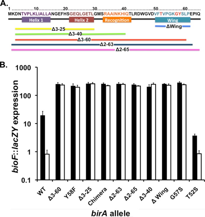
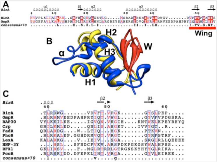
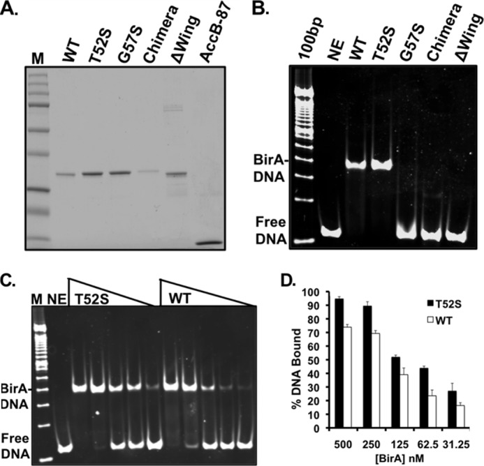
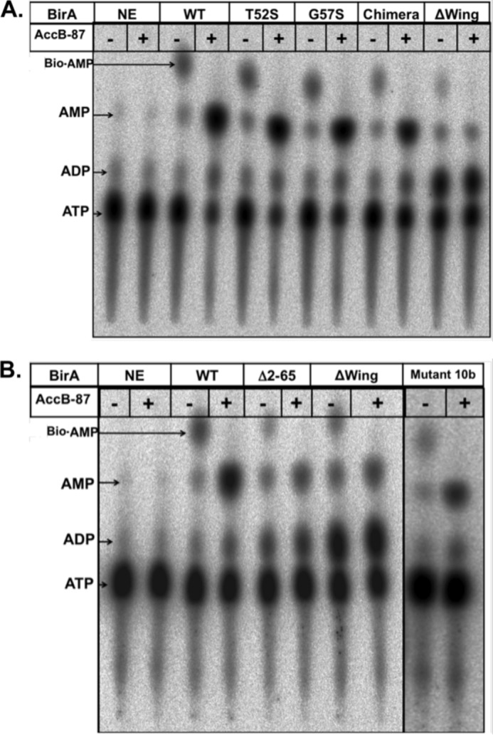
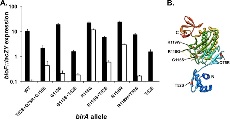
The BirA biotin protein ligase of Escherichia coli belongs to the winged helix-turn-helix (wHTH) family of transcriptional regulators. The N-terminal BirA domain is required for both transcriptional regulation of biotin synthesis and biotin protein ligase activity. We addressed the structural and functional role of the wing of the wHTH motif in both BirA functions. A panel of N-terminal deletion mutant proteins including a discrete deletion of the wing motif were unable to bind DNA. However, all the N-terminal deletion mutants weakly complemented growth of a ΔbirA strain at low biotin concentrations, indicating compromised ligase activity. A wing domain chimera was constructed by replacing the BirA wing with the nearly isosteric wing of the E. coli OmpR transcription factor. Although this chimera BirA was defective in operator binding, it was much more efficient in complementation of a ΔbirA strain than was the wing-less protein. The enzymatic activities of the wing deletion and chimera proteins in the in vitro synthesis of biotinoyl-5'-AMP differed greatly. The wing deletion BirA accumulated an off pathway compound, ADP, whereas the chimera protein did not. Finally, we report that a single residue alteration in the wing bypasses the deleterious effects caused by mutations in the biotin-binding loop of the ligase active site. We believe that the role of the wing in the BirA enzymatic reaction is to orient the active site and thereby protect biotinoyl-5'-AMP from attack by solvent. This is the first evidence that the wing domain of a wHTH protein can play an important role in enzymatic activity.

Keywords: AMP; Bacterial Genetics; Bacterial Metabolism; Enzyme Catalysis; Enzyme Mutation; Microbiology; Promoters; Transcription Factors; Transcription Regulation; Transcription Target Genes.


Figures

FIGURE 1.
FIGURE 1.
The biotin protein ligase reaction, the E. coli biotin operon regulation, and the three-dimensional structure of monomeric BirA protein. A shows the BPL activity of BirA. B and C show the general model of bio operon regulation. Green ovals denote BirA, tailed blue ovals are AccB, black dots represent biotin, and black dots with red pentagons denote biotinoyl-adenylate (Bio-5′-AMP). B shows the transcriptionally repressed state, whereas C shows the two modes of derepression of bio operon transcription engendered by either biotin limitation or excess unbiotinylated AccB acceptor protein. D shows the most recent BirA structure (Protein Data Bank 1HXD) in which the protein is complexed with biotinol-5′-AMP, an ester analog of Bio-5′-AMP (10). The N and C termini are denoted by N and C, respectively. Note that some loops were not visible in the crystals because of their high mobility and are shown as breaks in the chain. The figure was modified from Ref. .
FIGURE 2.
FIGURE 2.
Properties of the BirA N-terminal deletion and point mutant proteins. A, location of the mutants studied. The colored bars show the extents of the various deletions. The BirA residues have the same color code as the helix (the recognition helix is helix 3) except for the wing structure where the residues altered by point mutations are given in red type. B, expression of the bio operon in cultures grown with 10 nm biotin (derepression conditions; solid bars) or 10 μm (repression conditions; open bars) biotin assayed by use of β-galactosidase encoded by a chromosomal bioF::lacZY fusion. The strains used were derivatives of strain VC618 cured of plasmid pVC18 (8). Single colonies were streaked on defined medium plates supplemented with 10 nm biotin and X-gal for phenotypic confirmation. Liquid cultures grown in the same medium lacking X-gal and were then assayed for bio operon transcription by β-galactosidase activity (“Experimental Procedures”). Note that the data are plotted on the y axis as a log scale.
FIGURE 3.
FIGURE 3.
Complementation of the E. coli ΔbirA bioF::lacZY birA strain by expression of WT BirA, BirA T52S mutant protein, and BirA N-terminal deletion mutant proteins. The strains were assayed for growth in defined medium liquid cultures supplemented with various concentrations of biotin as given by the colored symbols of the third row as described under “Experimental Procedures.” Each of the panels is the host strain expressing a different birA allele. Each growth assay was carried out in triplicate, and this plot shows the averages of the triplicate cultures. Growth was measured by optical density at 600 nm. Note that the data are plotted on the y axis as a log scale. All the panels share the same color code. Expression was driven by the constitutive promoter of a medium copy number plasmid.
FIGURE 4.
FIGURE 4.
Sequences and structural alignments of the DNA-binding domains of several wHTH regulatory proteins. A, alignment of the C-terminal DNA-binding domain of OmpR with the N-terminal BirA DNA-binding domain. B, superimposition of the OmpR DNA-binding domain on the BirA N-terminal domain. The model was made using the Match Align Tool of Chimera (28) with Protein Data Bank files 1HXD and 1OPC. C, alignments of the wing motifs for several wHTH regulatory proteins of known structure were generated using the method described under “Experimental Procedures.” The Protein Data Bank files used were: BirA, 1HXD; OmpR, 1OPC; Rap30, 1BBY; CRP, 1CGP; FadR, 1E2X; PhoB, 1QQI; LexA, 1JHF; HNF-3γ, 1VTN; RFX1, 1DP7; and PcoR, 2JZY. Identical residues are denoted by white letters on a red background, similar residues are denoted by red letters on a white background, variable residues are in black letters, and dots represent gaps. In A and C, the alignment was generated using the BirA secondary structure as the reference structure. In A and C, the secondary structures of the proteins are shown at the top of the panel: β, β sheets; coils, β-turns; α, α-helices. In B, H1, H2, and H3 denote helices 1, 2, and 3, respectively. W denotes the wing structure, whereas α denotes the OmpR loop that interacts with RNA polymerase subunit α. Helix 3 is the recognition helix.
FIGURE 5.
FIGURE 5.
Purification and operator binding of purified BirA proteins. A, the BirA and AccB-87 proteins were subjected to electrophoresis in a 12% polyacrylamide gel. The Precision Plus protein standards from Bio-Rad are shown in lane M, and AccB-87 is in the far right-hand lane. The proteins were purified as described under “Experimental Procedures” and dialyzed in storage buffer prior to storage at −80 °C. In B, at 1 μm, the BirA mutant derivatives and wild type BirA purified as described under “Experimental Procedures” were assayed for binding to a 112-bp bioO duplex DNA prepared as described under “Experimental Procedures.” The reaction products were separated by native gel electrophoresis, and the gels were stained with SYBR Green I nucleic acid gel stain and visualized using the Bio-Rad Chemi Doc XRS. In C, the point mutant T52S and wild type BirA proteins (0–500 nm) were subjected to the same analysis. In D, the results from three independent experiments were quantified with the Quantity One program. % DNA bound represents the percentage of DNA bound to BirA protein versus the percentage of remaining unbound calculated for each lane using the no enzyme (NE) lane as control for the amount of DNA loaded. The results are presented as the averages and standard errors of triplicate determinations. The open bars represent the wild type BirA, and the closed bars represent the T52S mutant protein. The identities of the BirA proteins tested are given at the tops of A, B, and C. The standards in B and C are the 100-bp DNA ladder (Life Technologies).
FIGURE 6.
FIGURE 6.
Thin layer chromatographic analysis of various BirA proteins to synthesize Bio-5′-AMP and transfer the biotin moiety to AccB-87. Synthesis of biotinoyl-adenylate in reaction mixtures containing 1 μm BirA, 20 μm biotin, 5 μm ATP, 5 mm MgCl2, 100 mm KCl, 5 mm TCEP, and 16.5 nm [α-32P]ATP is shown. Each reaction was run in duplicate and incubated for 30 min at 37 °C, after which time point plus signs (+) purified apo-AccB-87 (50 μm) was added to one of the duplicate reactions followed by incubation for an additional 15 min. The second duplicate reaction did not receive AccB-87 (minus signs). One μl of each reaction was spotted onto a cellulose thin layer chromatography plate. The right panels with 10b BirA mutant show a separate experiment. Following plate development, the reaction products (indicated by arrows), biotinoyl-5′-AMP (Bio-AMP), ADP, AMP, and the remaining ATP were visualized by autoradiography. NE denotes the control reactions lacking BirA. The proteins assayed are given at the top of the figure, and the identities of the spots are on the left margin. A and B show the products of the BirA reaction in the presence or absence of the AccB-87 acceptor protein. The identity of each protein is given at the top of each pair of lanes. The wild type (WT) and Δwing proteins were included in both analyses as controls.
FIGURE 7.
FIGURE 7.
Genetic evidence that the wing plays a direct role in the enzymatic function of BirA. A, transcription of the bio operon in cultures grown with 4 nm biotin (derepression conditions; depicted with solid bars) or 4 μm biotin (repression conditions; depicted by open bars) assayed by β-galactosidase production from a chromosomal bioF::lacZY fusion. The strains used were derivatives of strain VC618 cured of plasmid pVC18 (8). Single colonies were streaked on defined medium plates supplemented with 4 nm biotin and X-gal for phenotypic confirmation. Liquid cultures were grown in the same medium lacking X-gal and were then assayed for bio operon transcription by β-galactosidase activity (“Experimental Procedures”). Note that the data are plotted on the y axis as a log scale. B, three-dimensional structure of monomeric BirA protein. The single residue substitutions tested are highlighted. The model was constructed using Chimera and Protein Data Bank file 1HXD.

References

    1. Beckett D. (2007) Biotin sensing. Universal influence of biotin status on transcription. Annu. Rev. Genet. 41, 443–464 - PubMed
    1. Cronan J. E., Jr. (1988) Expression of the biotin biosynthetic operon of Escherichia coli is regulated by the rate of protein biotination. J. Biol. Chem. 263, 10332–10336 - PubMed
    1. Streaker E. D., Gupta A., Beckett D. (2002) The biotin repressor. Thermodynamic coupling of corepressor binding, protein assembly, and sequence-specific DNA binding. Biochemistry 41, 14263–14271 - PubMed
    1. Mukhopadhyay B., Purwantini E., Kreder C. L., Wolfe R. S. (2001) Oxaloacetate synthesis in the methanarchaeon Methanosarcina barkeri. Pyruvate carboxylase genes and a putative Escherichia coli-type bifunctional biotin protein ligase gene (bpl/birA) exhibit a unique organization. J. Bacteriol. 183, 3804–3810 - PMC - PubMed
    1. Streaker E. D., Beckett D. (1998) A map of the biotin repressor-biotin operator interface. Binding of a winged helix-turn-helix protein dimer to a forty base-pair site. J. Mol. Biol. 278, 787–800 - PubMed

Publication types

MeSH terms

LinkOut - more resources