

Modeling pathologic response of esophageal cancer to chemoradiation therapy using spatial-temporal 18F-FDG PET features, clinical parameters, and demographics
- PMID: 24189128
- PMCID: PMC3875172
- DOI: 10.1016/j.ijrobp.2013.09.037
Modeling pathologic response of esophageal cancer to chemoradiation therapy using spatial-temporal 18F-FDG PET features, clinical parameters, and demographics
Abstract
Purpose: To construct predictive models using comprehensive tumor features for the evaluation of tumor response to neoadjuvant chemoradiation therapy (CRT) in patients with esophageal cancer.
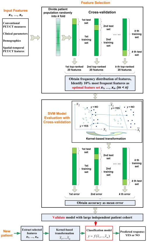
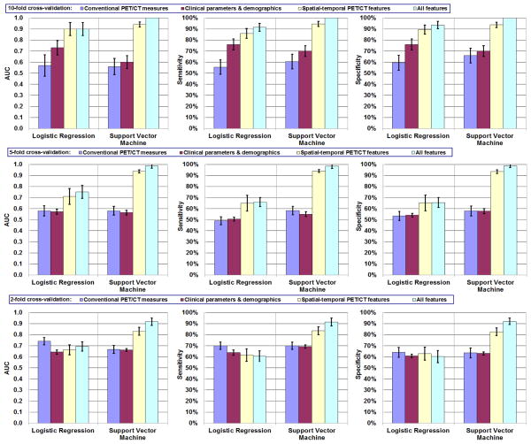
Methods and materials: This study included 20 patients who underwent trimodality therapy (CRT+surgery) and underwent 18F-fluorodeoxyglucose (FDG) positron emission tomography/computed tomography (PET/CT) both before and after CRT. Four groups of tumor features were examined: (1) conventional PET/CT response measures (eg, standardized uptake value [SUV]max, tumor diameter); (2) clinical parameters (eg, TNM stage, histology) and demographics; (3) spatial-temporal PET features, which characterize tumor SUV intensity distribution, spatial patterns, geometry, and associated changes resulting from CRT; and (4) all features combined. An optimal feature set was identified with recursive feature selection and cross-validations. Support vector machine (SVM) and logistic regression (LR) models were constructed for prediction of pathologic tumor response to CRT, cross-validations being used to avoid model overfitting. Prediction accuracy was assessed by area under the receiver operating characteristic curve (AUC), and precision was evaluated by confidence intervals (CIs) of AUC.
Results: When applied to the 4 groups of tumor features, the LR model achieved AUCs (95% CI) of 0.57 (0.10), 0.73 (0.07), 0.90 (0.06), and 0.90 (0.06). The SVM model achieved AUCs (95% CI) of 0.56 (0.07), 0.60 (0.06), 0.94 (0.02), and 1.00 (no misclassifications). With the use of spatial-temporal PET features combined with conventional PET/CT measures and clinical parameters, the SVM model achieved very high accuracy (AUC 1.00) and precision (no misclassifications)-results that were significantly better than when conventional PET/CT measures or clinical parameters and demographics alone were used. For groups with many tumor features (groups 3 and 4), the SVM model achieved significantly higher accuracy than did the LR model.
Conclusions: The SVM model that used all features including spatial-temporal PET features accurately and precisely predicted pathologic tumor response to CRT in esophageal cancer.
Copyright © 2014 Elsevier Inc. All rights reserved.
Conflict of interest statement
Figures
Similar articles
-
Spatial-temporal [¹⁸F]FDG-PET features for predicting pathologic response of esophageal cancer to neoadjuvant chemoradiation therapy.Int J Radiat Oncol Biol Phys. 2013 Apr 1;85(5):1375-82. doi: 10.1016/j.ijrobp.2012.10.017. Epub 2012 Dec 6. Int J Radiat Oncol Biol Phys. 2013. PMID: 23219566 Free PMC article.
-
The role of qualitative and quantitative analysis of F18-FDG positron emission tomography in predicting pathologic response following chemoradiotherapy in patients with esophageal carcinoma.J Gastrointest Cancer. 2012 Dec;43(4):612-8. doi: 10.1007/s12029-012-9412-3. J Gastrointest Cancer. 2012. PMID: 22777832
-
2-Fluoro-2-deoxy-D-glucose positron emission tomography imaging is predictive of pathologic response and survival after preoperative chemoradiation in patients with esophageal carcinoma.Cancer. 2004 Oct 15;101(8):1776-85. doi: 10.1002/cncr.20585. Cancer. 2004. PMID: 15386332
-
Utility of PET, CT, and EUS to identify pathologic responders in esophageal cancer.Ann Thorac Surg. 2004 Oct;78(4):1152-60; discussion 1152-60. doi: 10.1016/j.athoracsur.2004.04.046. Ann Thorac Surg. 2004. PMID: 15464463 Review.
-
Role of positron emission tomography in the (re-)staging of oesophageal cancer.Scand J Gastroenterol Suppl. 2006;(243):116-22. doi: 10.1080/00365520600664409. Scand J Gastroenterol Suppl. 2006. PMID: 16782630 Review.
Cited by
-
Preoperative 18F-FDG PET/CT and CT radiomics for identifying aggressive histopathological subtypes in early stage lung adenocarcinoma.Comput Struct Biotechnol J. 2023 Nov 4;21:5601-5608. doi: 10.1016/j.csbj.2023.11.008. eCollection 2023. Comput Struct Biotechnol J. 2023. PMID: 38034400 Free PMC article.
-
Radiomics in medical imaging: pitfalls and challenges in clinical management.Jpn J Radiol. 2022 Sep;40(9):919-929. doi: 10.1007/s11604-022-01271-4. Epub 2022 Mar 28. Jpn J Radiol. 2022. PMID: 35344132 Review.
-
The emerging field of radiomics in esophageal cancer: current evidence and future potential.Transl Cancer Res. 2016 Aug;5(4):410-423. doi: 10.21037/tcr.2016.06.19. Transl Cancer Res. 2016. PMID: 30687593 Free PMC article.
-
Predicting the Response of Neoadjuvant Therapy for Patients with Esophageal Carcinoma: an In-depth Literature Review.J Cancer. 2015 Sep 15;6(11):1179-86. doi: 10.7150/jca.12346. eCollection 2015. J Cancer. 2015. PMID: 26516367 Free PMC article. Review.
-
Editorial on "Can CT-PET and endoscopic assessment post-neoadjuvant chemoradiotherapy predict residual disease in esophageal cancer".J Thorac Dis. 2017 Oct;9(10):3645-3648. doi: 10.21037/jtd.2017.09.117. J Thorac Dis. 2017. PMID: 29268364 Free PMC article. No abstract available.
References
-
- American Cancer Society. [Accessed on May 13, 2013..];Cancer Facts & Figures. 2012 Available from: www.cancer.org/Research/CancerFactsFigures/CancerFactsFigures/cancer-fac....
-
- Stahl M, Stuschke M, Lehmann N, et al. Chemoradiation with and without surgery in patients with locally advanced squamous cell carcinoma of the esophagus. Journal of Clinical Oncology. 2005;23:2310–2317. - PubMed
-
- Bedenne L, Michel P, Bouche O, et al. Chemoradiation followed by surgery compared with chemoradiation alone in squamous cancer of the esophagus: FFCD 9102. J Clin Oncol. 2007;25:1160–1168. - PubMed
Publication types
MeSH terms
Substances
Grants and funding
LinkOut - more resources
Full Text Sources
Other Literature Sources
Medical
Research Materials
