. 2014 Jan;92(1):90-8.
doi: 10.1038/icb.2013.70. Epub 2013 Nov 5.

Colitogenic effector T cells: roles of gut-homing integrin, gut antigen specificity and γδ T cells

Affiliations

Colitogenic effector T cells: roles of gut-homing integrin, gut antigen specificity and γδ T cells

Jeong-Su Do et al. Immunol Cell Biol. 2014 Jan.

Abstract

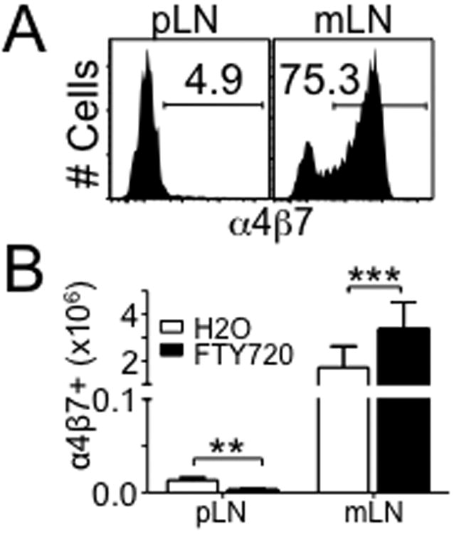
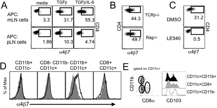
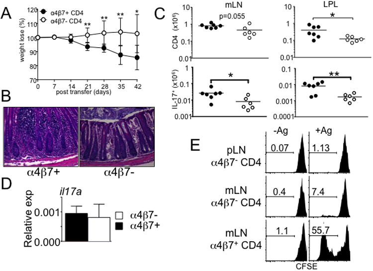
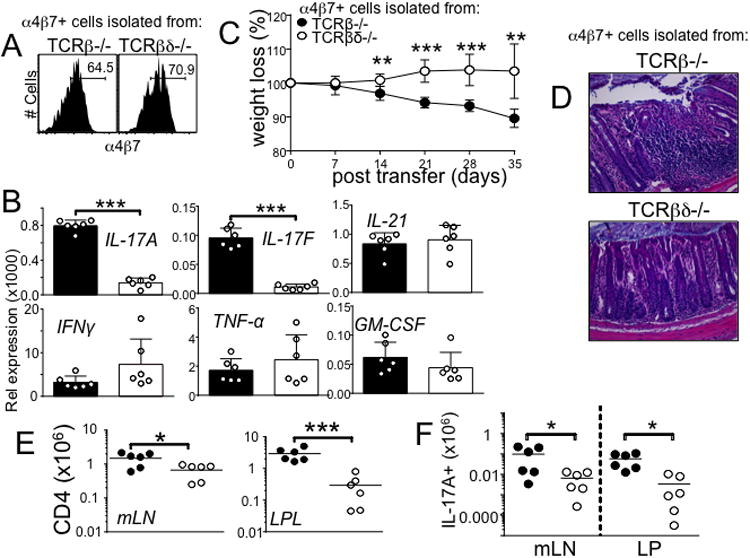
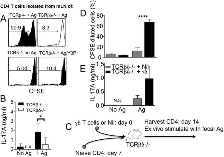
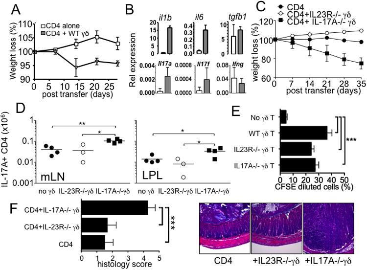
Disturbance of T-cell homeostasis could lead to intestinal inflammation. Naive CD4 T cells undergoing spontaneous proliferation, a robust proliferative response that occurs under severe lymphopenic conditions, differentiate into effector cells producing Th1- and/or Th17-type cytokines and induce a chronic inflammation in the intestine that resembles human inflammatory bowel disease. In this study, we investigated the key properties of CD4 T cells necessary to induce experimental colitis. α4β7 upregulation was primarily induced by mesenteric lymph node (mLN) resident CD11b(+) dendritic cell subsets via transforming growth factor beta (TGFβ)/retinoic acid-dependent mechanism. Interestingly, α4β7 expression was essential but not sufficient to induce inflammation. In addition to gut-homing specificity, expression of gut Ag specificity was also crucial. T-cell acquisition of the specificity was dramatically enhanced by the presence of γδ T cells, a population previously shown to exacerbate T-cell-mediated colitis. Importantly, interleukin (IL)-23-mediated γδ T cell stimulation was necessary to enhance colitogenicity but not gut antigen reactivity of proliferating CD4 T cells. These findings demonstrate that T-cell colitogenicity is achieved through multiple processes, offering a therapeutic rationale by intervening these pathways.


Figures

Figure 1
Figure 1. α4β7 expression on CD4 T cells was enhanced in mesenteric lymph node
(A) α4β7 expression on CD4 T cells at day 5 following CD4 T cells adoptive transfer to TCRβ-/- mice. Data shown are representative of individually tested recipients. All experiments were repeated more than three times and similar results were observed. (B) Total number of α4β7+ CD4 T cells from the indicated organ after FTY720 treatment. Data shown are the mean ± SD of 6 mice in two independent experiments. **, p<0.01; ***, p<0.001.
Figure 2
Figure 2. mLN CD11c+ CD11b+ cells induced α4β7 expression on T cells dependent on retinoic acid
(A) OVA-specific OT-II T cells were cocultured with cells from the indicated tissues in the presence of TGFβ and IL-6. α4β7 expression on OT-II cells was measured after 3 days of culture. The experiments were repeated five times and similar results were observed. (B) OT-II T cells were stimulated with mLN cells from TCRβ-/- or Rag-/- mice. α4β7 expression on OT-II cells was determined. Plots are representative of at least three independent experiments. (C) OT-II cell/mLN cell coculture was repeated in the presence of LE540 or control vehicle. α4β7 expression was similarly measured as above. The experiments were repeated three times and similar results were observed. (D) mLN cells of the indicated phenotypes were FACS sorted and cocultured with OT-II T cells in the presence of Ag. Filled histogram represents α4β7 expression without Ag. The experiments were repeated twice and similar results were observed. *, p<0.05; **, p<0.01. (E) CD103 expression on mLN DC subsets. Data are representative of at least three independent experiments.
Figure 3
Figure 3. More severe inflammation develops in TCRβ-/- mice by α4β7+ CD4 T cells compare to α4β7 CD4 T cells
Naïve CD4 T cells were transferred into TCRβ-/- mice. α4β7+ and α4β7 donor T cells were isolated from the mLN 3 weeks post transfer, and retransferred into naïve TCRβ-/- recipients. (A) Weight loss and (B) colon histology in TCRβ-/- mice at 6 weeks post-transfer of α4β7+ or α4β7 CD4 T cells. All images at 20× magnification. Data shown are the mean ± SD. Experiments were repeated twice and similar results were observed. *, p<0.05; **, p<0.01. (C) Absolute number of the total cells and IL-17-producing donor T cells from the indicated organ are shown. Data are mean ± SD of 6-7 mice from two independent experiments. *, p<0.05; **, p<0.01. (D) Real time quantitative PCR analysis of il17a expression on α4β7+ and α4β7 CD4 T cells prior to the transfer. All samples were done in duplicates and normalized to GAPDH. Data shown are representative from 6-7 individually tested mice. (E) α4β7+ and α4β7 donor T cells were reisolated from the mLN and pLN, CFSE labeled, and cocultured with APCs pulsed with fecal extract Ag as described in Methods. CFSE dilution was determined by FACS analysis. Experiments were repeated twice, and similar results were obtained.
Figure 4
Figure 4. α4β7+ CD4 cells from TCRβ-/- mice display severe colitogenicity compared to α4β7+ CD4 cells from TCRβδ-/- mice
(A) α4β7 expression on CD4 T cells at day 21 following CD4 T cells adoptive transfer to TCRβ-/- and TCRβδ-/- mice. Data shown are representative from 6 individually tested mice in two independent experiments. (B) Real time quantitative PCR analysis of pro-inflammatory gene expression of α4β7+ CD4 T cells. The expression was normalized to GAPDH. Data shown are representative from 6 individually tested mice. ***, p<0.001. (C) Weight loss and (D) colon histology in TCRβ-/- recipient mice at 5 weeks after post-transfer of α4β7+ CD4 T cells from TCRβ-/- or TCRβδ-/- mice. All images at 20× magnification. Data are representative of two independent experiments. **, p<0.01; ***, p<0.001. (E) Absolute number of total cells and (F) IL-17-producing cells from the indicated tissues. Data are mean ± SD of 6 individually tested mice. *, p<0.05; ***, p<0.001.
Figure 5
Figure 5. α4β7+ CD4 T cells from TCRβ-/- recipients strongly respond to fecal extract antigen stimulation ex vivo
(A) Following naïve CD4 T cell transfer into TCRβ-/- or TCRβδ-/- mice, α4β7+ CD4 cells were isolated from individual recipient mice, CFSE labeled, and cocultured with fecal extract Ag pulsed APCs. CFSE dilution was analyzed at day 5 after stimulation. (B) IL-17 production in the culture supernatant was determined by ELISA. Data are shown of 7-12 individually tested mice in three independent experiments. (C) Experimental model. γδ T cells isolated from the lymphoid tissues were transferred into TCRβδ-/- mice. Seven days later, FACS sorted naïve CD4 T cells were transferred into the same recipients. The donor CD4 T cells were isolated from the mLN 7 days post T cell transfer, and cocultured with fecal extract Ag pulsed APCs. (D) CFSE dilution and (E) IL-17 production in the culture supernatant were determined. Experiments were repeated twice, and similar results were obtained. *, p<0.05; ****, p<0.0001.
Figure 6
Figure 6. IL-23-dependent stimulation of γδ T cells is required to enhance colitogenic CD4 T cell development
(A and C) FACS sorted CD4 T cells and γδ T cells (wild type, IL-23R-/-, and IL-17A-/-) were cotransferred into TCRβδ-/- mice, and weight loss was weekly monitored. (B) Expression of the indicated genes from CD4 alone (open bar) and CD4 plus wild type γδ T cells (grey bar) was determined by real time PCR analysis. (D) IL-17-producing donor CD4 T cells in the indicated tissues were determined 5 weeks post transfer. (E) Wild type, IL-23R-/-, and IL-17A-/- γδ T cells were transferred into TCRβδ-/- mice followed by naïve CD4 T cell transfer 7 days later as described in Fig 5. Donor CD4 T cells were isolated from the recipients, and cocultured with fecal extract Ag pulsed APCs as described in Fig 5. Experiments were repeated more than twice with similar results. (F) The recipients described above were sacrificed and the colon tissues were H&E stained for histopathology. The colitis score was measured as described in Methods. *, p<0.05; **, p<0.01; ***, p<0.001.

References

    1. Powrie F, Leach MW, Mauze S, Menon S, Caddle LB, Coffman RL. Inhibition of Th1 responses prevents inflammatory bowel disease in scid mice reconstituted with CD45RBhi CD4+ T cells. Immunity. 1994;1:553–62. - PubMed
    1. Ostanin DV, Bao J, Koboziev I, Gray L, Robinson-Jackson SA, Kosloski-Davidson M, et al. T cell transfer model of chronic colitis: concepts, considerations, and tricks of the trade. Am J Physiol Gastrointest Liver Physiol. 2009;296:G135–46. - PMC - PubMed
    1. Do JS, Visperas A, Dong C, Baldwin WM, 3rd, Min B. Cutting edge: Generation of colitogenic Th17 CD4 T cells is enhanced by IL-17+ gammadelta T cells. J Immunol. 2011;186:4546–50. - PMC - PubMed
    1. Kieper WC, Troy A, Burghardt JT, Ramsey C, Lee JY, Jiang HQ, et al. Recent immune status determines the source of antigens that drive homeostatic T cell expansion. J Immunol. 2005;174:3158–63. - PubMed
    1. Johansson-Lindbom B, Agace WW. Generation of gut-homing T cells and their localization to the small intestinal mucosa. Immunol Rev. 2007;215:226–42. - PubMed

Publication types

MeSH terms