

Application of DNA chip scanning technology for automatic detection of Chlamydia trachomatis and Chlamydia pneumoniae inclusions
- PMID: 24189259
- PMCID: PMC3910709
- DOI: 10.1128/AAC.01400-13
Application of DNA chip scanning technology for automatic detection of Chlamydia trachomatis and Chlamydia pneumoniae inclusions
Abstract
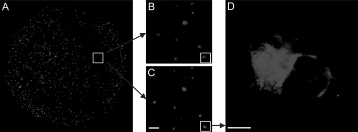
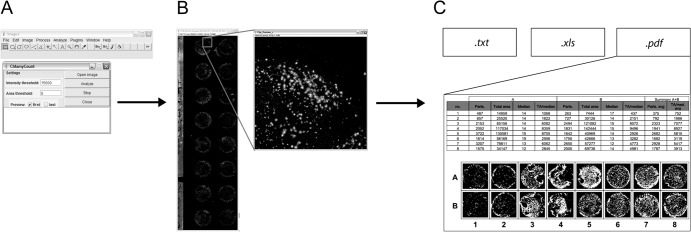
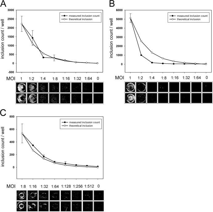
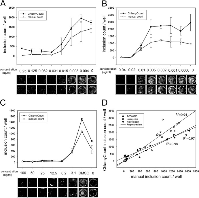
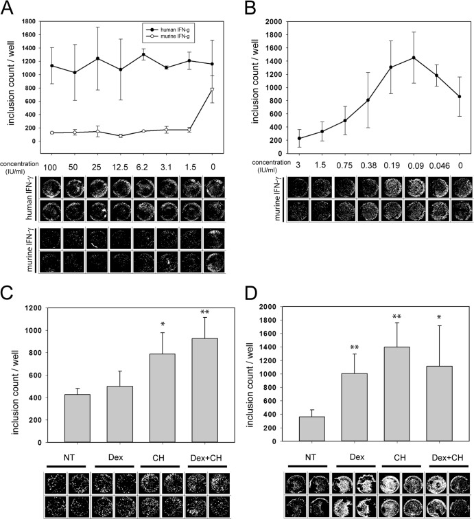
Chlamydiae are obligate intracellular bacteria that propagate in the inclusion, a specific niche inside the host cell. The standard method for counting chlamydiae is immunofluorescent staining and manual counting of chlamydial inclusions. High- or medium-throughput estimation of the reduction in chlamydial inclusions should be the basis of testing antichlamydial compounds and other drugs that positively or negatively influence chlamydial growth, yet low-throughput manual counting is the common approach. To overcome the time-consuming and subjective manual counting, we developed an automatic inclusion-counting system based on a commercially available DNA chip scanner. Fluorescently labeled inclusions are detected by the scanner, and the image is processed by ChlamyCount, a custom plug-in of the ImageJ software environment. ChlamyCount was able to measure the inclusion counts over a 1-log-unit dynamic range with a high correlation to the theoretical counts. ChlamyCount was capable of accurately determining the MICs of the novel antimicrobial compound PCC00213 and the already known antichlamydial antibiotics moxifloxacin and tetracycline. ChlamyCount was also able to measure the chlamydial growth-altering effect of drugs that influence host-bacterium interaction, such as gamma interferon, DEAE-dextran, and cycloheximide. ChlamyCount is an easily adaptable system for testing antichlamydial antimicrobials and other compounds that influence Chlamydia-host interactions.
Figures
Similar articles
-
High dynamic range detection of Chlamydia trachomatis growth by direct quantitative PCR of the infected cells.J Microbiol Methods. 2016 Jan;120:15-22. doi: 10.1016/j.mimet.2015.11.010. Epub 2015 Nov 11. J Microbiol Methods. 2016. PMID: 26578244
-
Establishment of a particle-counting method for purified elementary bodies of chlamydiae and evaluation of sensitivities of the IDEIA Chlamydia kit and DNA probe by using the purified elementary bodies.J Clin Microbiol. 1992 Nov;30(11):2911-6. doi: 10.1128/jcm.30.11.2911-2916.1992. J Clin Microbiol. 1992. PMID: 1452662 Free PMC article.
-
Rab GTPases are recruited to chlamydial inclusions in both a species-dependent and species-independent manner.Infect Immun. 2003 Oct;71(10):5855-70. doi: 10.1128/IAI.71.10.5855-5870.2003. Infect Immun. 2003. PMID: 14500507 Free PMC article.
-
[Diagnostic tests: Chlamydia trachomatis, Chlamydophila pneumoniae].Nihon Rinsho. 2005 Jul;63 Suppl 7:220-6. Nihon Rinsho. 2005. PMID: 16111232 Review. Japanese. No abstract available.
-
[Effector proteins of Clamidia].Mol Biol (Mosk). 2009 Nov-Dec;43(6):963-83. Mol Biol (Mosk). 2009. PMID: 20088373 Review. Russian.
Cited by
-
Efficacy of two mouth rinse sprays in inhibiting Streptococcus mutans growth on toothbrush bristles.Saudi Dent J. 2018 Oct;30(4):365-372. doi: 10.1016/j.sdentj.2018.07.005. Epub 2018 Aug 4. Saudi Dent J. 2018. PMID: 30202175 Free PMC article.
-
Chlamydia pneumoniae Infection Exacerbates Atherosclerosis in ApoB100only/LDLR-/- Mouse Strain.Biomed Res Int. 2018 Mar 25;2018:8325915. doi: 10.1155/2018/8325915. eCollection 2018. Biomed Res Int. 2018. PMID: 29770337 Free PMC article.
-
Selenocompounds as Novel Antibacterial Agents and Bacterial Efflux Pump Inhibitors.Molecules. 2019 Apr 16;24(8):1487. doi: 10.3390/molecules24081487. Molecules. 2019. PMID: 31014009 Free PMC article.
-
Alternative strategies for Chlamydia treatment: Promising non-antibiotic approaches.Front Microbiol. 2022 Nov 23;13:987662. doi: 10.3389/fmicb.2022.987662. eCollection 2022. Front Microbiol. 2022. PMID: 36504792 Free PMC article. Review.
-
Vaginal Gel Component Hydroxyethyl Cellulose Significantly Enhances the Infectivity of Chlamydia trachomatis Serovars D and E.Antimicrob Agents Chemother. 2018 Dec 21;63(1):e02034-18. doi: 10.1128/AAC.02034-18. Print 2019 Jan. Antimicrob Agents Chemother. 2018. PMID: 30373805 Free PMC article.
References
-
- Mishori R, McClaskey EL, WinklerPrins VJ. 2012. Chlamydia trachomatis infections: screening, diagnosis, and management. Am. Fam. Physician 86:1127–1132 - PubMed
Publication types
MeSH terms
LinkOut - more resources
Full Text Sources
Other Literature Sources
