. 2013 Nov 11;203(3):417-25.
doi: 10.1083/jcb.201303071. Epub 2013 Nov 4.

Two appendages homologous between basal bodies and centrioles are formed using distinct Odf2 domains

Affiliations

Two appendages homologous between basal bodies and centrioles are formed using distinct Odf2 domains

Kazuhiro Tateishi et al. J Cell Biol. .

Abstract

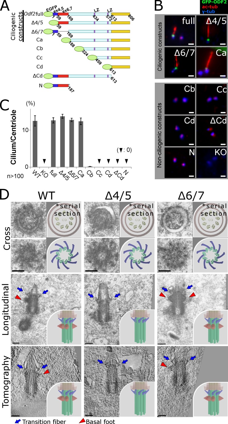
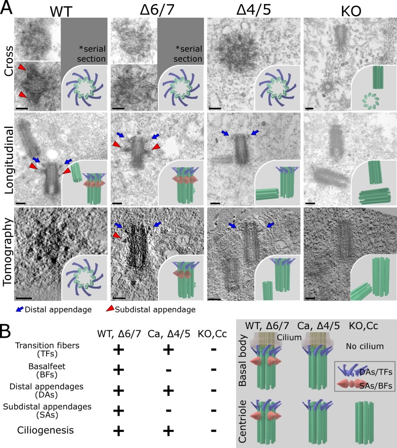
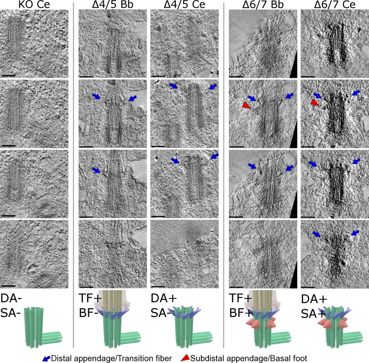
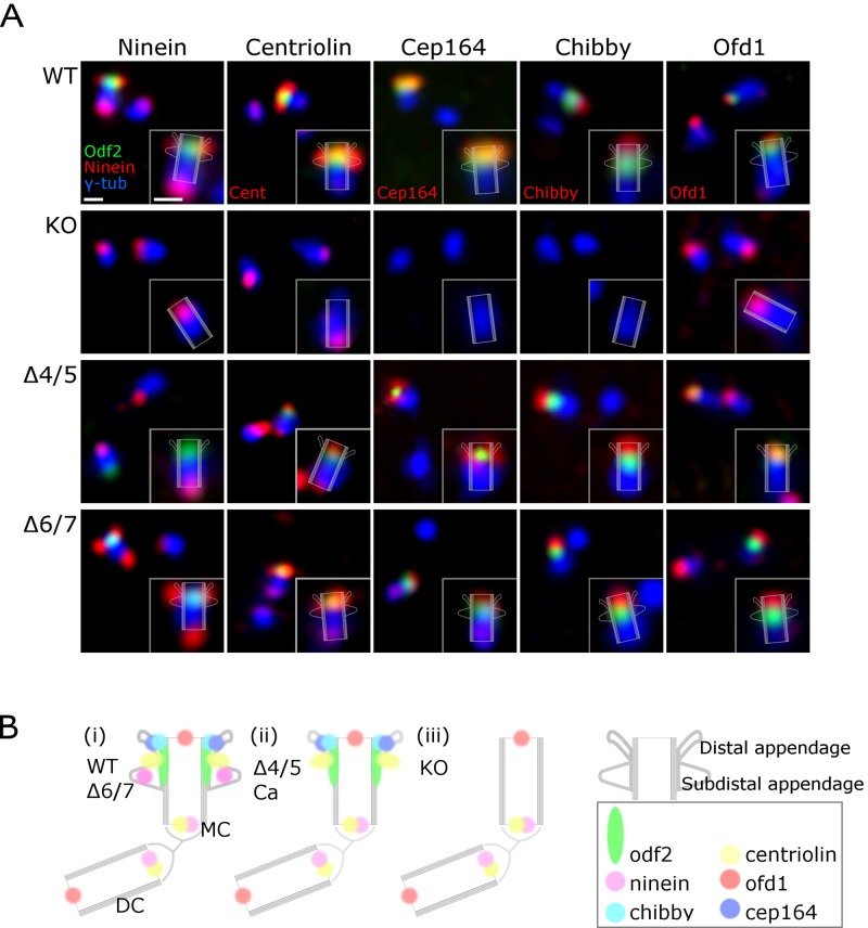
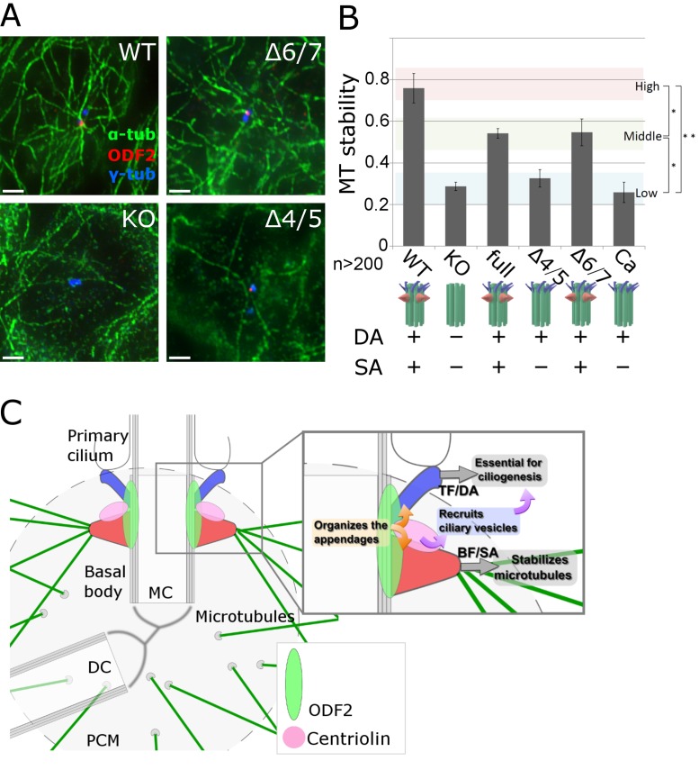
Ciliogenesis is regulated by context-dependent cellular cues, including some transduced through appendage-like structures on ciliary basal bodies called transition fibers and basal feet. However, the molecular basis for this regulation is not fully understood. The Odf2 gene product, ODF2/cenexin, is essential for both ciliogenesis and the formation of the distal and subdistal appendages on centrioles, which become basal bodies. We examined the effects of Odf2 deletion constructs on ciliogenesis in Odf2-knockout F9 cells. Electron microscopy revealed that ciliogenesis and transition fiber formation required the ODF2/cenexin fragment containing amino acids (aa) 188-806, whereas basal foot formation required aa 1-59 and 188-806. These sequences also formed distal and subdistal appendages, respectively, indicating that the centriole appendages are molecularly analogous to those on basal bodies. We used the differential formation of appendages by Odf2 deletion constructs to study the incorporation and function of molecules associated with each appendage type. We found that transition fibers and distal appendages were required for ciliogenesis and subdistal appendages stabilized the centrosomal microtubules.


Figures

Figure 1.
Figure 1.
Odf2 gene sequences required to generate cilia and reconstitute the appendages of ciliary basal bodies. (A) Schematic representation of the Odf2 deletion constructs, which were transfected into Odf2-KO F9 cells. LZ, leucine zipper motif. (B) Immunofluorescence for GFP (Odf2), γ-tubulin (centrioles/basal bodies), and acetylated tubulin (primary cilia) to examine the generation of cilia. Bars, 1 µm. (C) Percentage of cilia on centrioles in cells expressing the indicated construct (n > 100 in more than three independent experiments). (D) Electron micrographs showing transition fibers (TFs) and/or basal feet (BF) on ciliary basal bodies. Thin-section electron microscopic images show cross sections (Cross) and longitudinal sections (Longitudinal) of primary cilia. Tomography, UHVEMT images of basal bodies; TFs, blue arrows; BF, red arrowheads; WT, (TF+BF+) basal bodies in WT F9 cells; Δ4/5, (TF+BF−) basal bodies in Δ4/5 construct–expressing Odf2-KO F9 cells; Δ6/7, (TF+BF+) basal bodies in Δ6/7 construct–expressing Odf2-KO F9 cells. Bars, 0.2 µm. More than five samples were analyzed in each case. The cross section electron micrographs in the top two rows are the same images as in the top two rows of Fig. S3 A, where they are annotated for TFs and BF. Insets, schematic drawings of electron microscopic images of basal bodies.
Figure 2.
Figure 2.
Reconstitution of centriole appendages by Odf2 deletion constructs. (A) Electron micrographs showing distal appendages (DAs) and/or subdistal appendages (SAs) on centrioles. Thin-section electron microscopic images show cross sections (Cross) and longitudinal sections (Longitudinal) of centrioles. More than five samples were analyzed in each case. Tomography, UHVEMT images of centrioles; DAs, blue arrows; SAs, red arrowheads; WT, (DA+SA+) centriole in WT F9 cells; Δ4/5, (DA+SA−) centriole in Δ4/5 construct–expressing Odf2-KO F9 cells; Δ6/7, (DA+SA+) centriole in Δ6/7 construct–expressing Odf2-KO F9 cells; KO, (DA−SA−) centriole; TFs, blue arrows; BF, red arrowheads. Bars, 0.2 µm. Insets, schematic drawings of electron microscopic images of centrioles. (B) Summary of findings on the reconstitution of the basal body/centriole appendages.
Figure 3.
Figure 3.
Ultra-high voltage electron microscopic tomographic (UHVEMT) images of basal bodies/centrioles. Four slices are shown (see Videos 1–9). Bb, basal body; Ce, centriole; Δ4/5 and Δ6/7, deletion mutants of Odf2 that were exogenously expressed in Odf2-KO F9 cells; TFs/DAs, blue arrows; BF/SAs, red arrowheads. Bars, 0.2 µm.
Figure 4.
Figure 4.
Immunofluorescence microscopic images of centriole appendages in the DA+SA+, DA-SA−, and DA+SA− patterns. (A) Immunofluorescence for ninein, centriolin, CEP164, and OFD1. WT, (DA+SA+) centriole in WT F9 cells; KO, (DA-SA−) centriole in Odf2-KO F9 cells; Δ4/5, (DA+SA−) centriole in Δ4/5 construct–expressing Odf2-KO F9 cells; Δ6/7, (DA+SA+) centriole in Δ6/7 construct–expressing Odf2-KO F9 cells. (B) Schematic drawing showing the localization of centrosomal components. MC, mother centriole; DC, daughter centriole. Bars, 500 nm.
Figure 5.
Figure 5.
Specific role of centrosomal subdistal appendages (SAs) in stabilizing centriole microtubules (MTs). (A) Immunofluorescence images of MTs in DA+SA+, DA+SA−, and DA−SA− centrioles under nocodazole treatment, an MT-destabilizing condition. WT, (DA+SA+) centriole in WT F9 cells; Δ4/5, (DA+SA−) centriole in Δ4/5 construct–expressing Odf2-KO F9 cells; Δ6/7, (DA+SA+) centriole in Δ6/7 construct–expressing Odf2-KO F9 cells. (B) Quantification of MT stability. The relative MT stability is shown for the indicated Odf2 construct (*, P = 0.01; **, P = 0.005; n > 200 in more than three independent experiments). (C) Schematic drawing of the specific roles of the appendages of ciliary basal bodies/centrioles. In the proposed model, separate domains in Odf2 serve as the molecular platform on which the appendages are constructed. Note that centriolin associates with ODF2/cenexin at the base of the SA (or where the base would be in the absence of SAs), and recruits vesicles whose cargoes support ciliogenesis. The BF/SAs (red) stabilize MTs, whereas the TFs/DAs (blue) are essential for ciliogenesis. MC, mother centriole; DC, daughter centriole.

Similar articles

Cited by

References

    1. Anderson R.G. 1972. The three-dimensional structure of the basal body from the rhesus monkey oviduct. J. Cell Biol. 54:246–265 10.1083/jcb.54.2.246 - DOI - PMC - PubMed
    1. Azimzadeh J., Marshall W.F. 2010. Building the centriole. Curr. Biol. 20:R816–R825 10.1016/j.cub.2010.08.010 - DOI - PMC - PubMed
    1. Bornens M. 2002. Centrosome composition and microtubule anchoring mechanisms. Curr. Opin. Cell Biol. 14:25–34 10.1016/S0955-0674(01)00290-3 - DOI - PubMed
    1. Bornens M., Paintrand M., Berges J., Marty M.C., Karsenti E. 1987. Structural and chemical characterization of isolated centrosomes. Cell Motil. Cytoskeleton. 8:238–249 10.1002/cm.970080305 - DOI - PubMed
    1. Bouckson-Castaing V., Moudjou M., Ferguson D.J., Mucklow S., Belkaid Y., Milon G., Crocker P.R. 1996. Molecular characterisation of ninein, a new coiled-coil protein of the centrosome. J. Cell Sci. 109:179–190 - PubMed

Publication types

LinkOut - more resources