. 2014 Jun;28(6):1308-15.
doi: 10.1038/leu.2013.326. Epub 2013 Nov 5.

CD133 is a positive marker for a distinct class of primitive human cord blood-derived CD34-negative hematopoietic stem cells

Affiliations
Free PMC article

CD133 is a positive marker for a distinct class of primitive human cord blood-derived CD34-negative hematopoietic stem cells

M Takahashi et al. Leukemia. 2014 Jun.
Free PMC article

Abstract

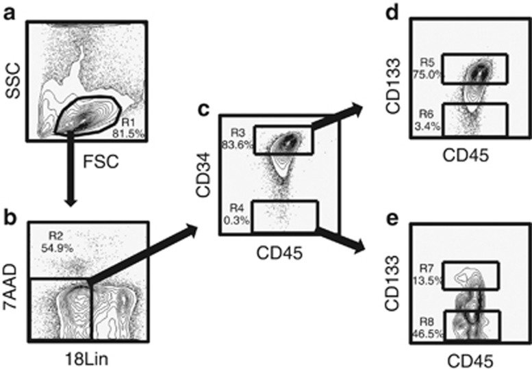
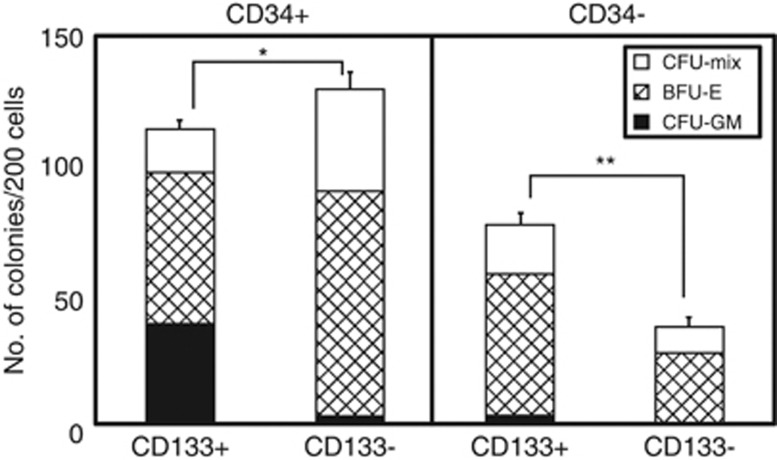
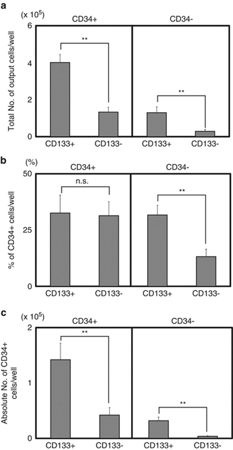
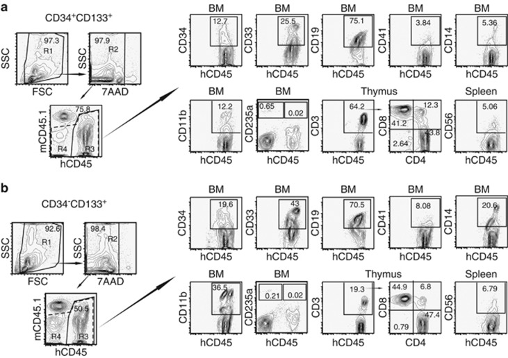
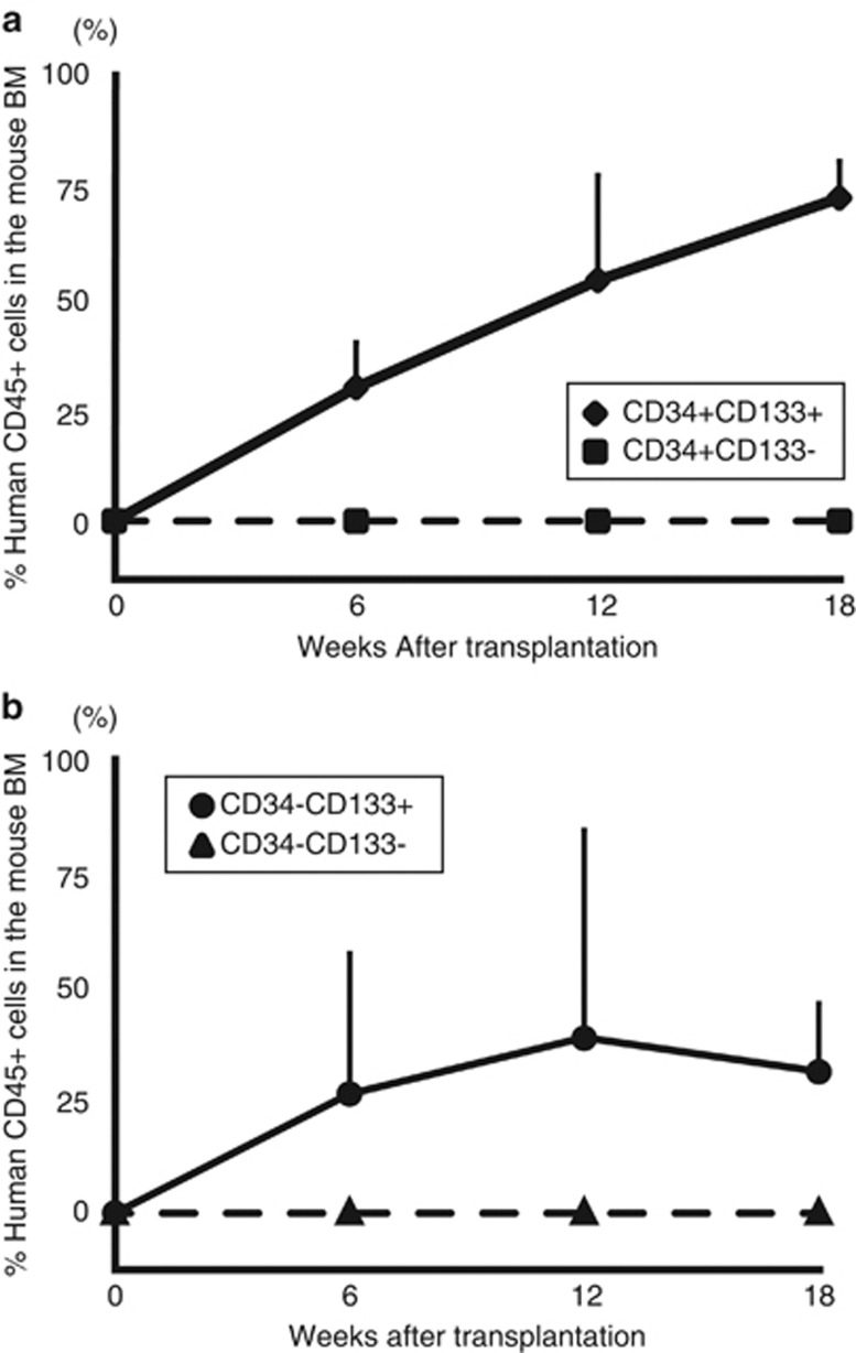
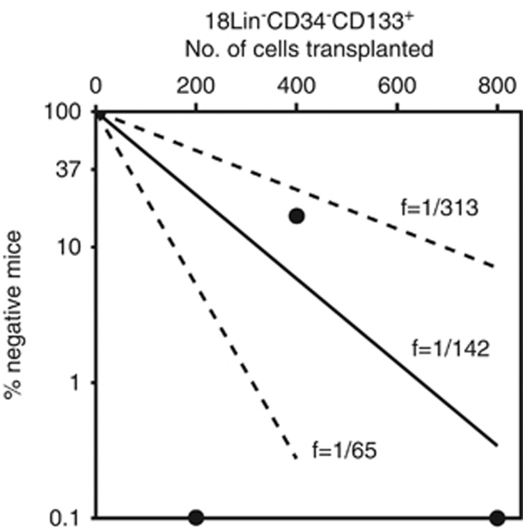
The identification of human CD34-negative (CD34(-)) hematopoietic stem cells (HSCs) provides a new concept for the hierarchy in the human HSC compartment. Previous studies demonstrated that CD34(-) severe combined immunodeficiency (SCID)-repopulating cells (SRCs) are a distinct class of primitive HSCs in comparison to the well-characterized CD34(+)CD38(-) SRCs. However, the purification level of rare CD34(-) SRCs in 18 lineage marker-negative (Lin(-)) CD34(-) cells (1/1000) is still very low compared with that of CD34(+)CD38(-) SRCs (1/40). As in the mouse, it will be necessary to identify useful positive markers for a high degree of purification of rare human CD34(-) SRCs. Using 18Lin(-)CD34(-) cells, we analyzed the expression of candidate positive markers by flow cytometric analysis. We finally identified CD133 as a reliable positive marker of human CB-derived CD34(-) SRCs and succeeded in highly purifying primitive human CD34(-) HSCs. The limiting dilution analysis demonstrated that the incidence of CD34(-) SRCs in 18Lin(-)CD34(-)CD133(+) cells was 1/142, which is the highest level of purification of these unique CD34(-) HSCs to date. Furthermore, CD133 expression clearly segregated the SRC activities of 18Lin(-)CD34(-) cells, as well as 18Lin(-)CD34(+) cells, in their positive fractions, indicating its functional significance as a common cell surface maker to isolate effectively both CD34(+) and CD34(-) SRCs.


Figures

Figure 1
Figure 1
Representative FACS profiles of CB-derived 18LinCD45+CD34+/−CD133+/− cells. (a) The FSC/SSC profile of immunomagnetically separated Lin cells. The R1 gate was set on the blast–lymphocyte window. (b) The R2 gate was set on the 18Lin living cells. (c) The 18Lin living cells (R2) were subdivided into two fractions: 18LinCD45+CD34+ (R3) and CD34 (R4) cells, according to their expression levels of CD34. The definitions of CD34+/− cells are as follows: the CD34+ fraction contains cells expressing maximum APC fluorescence intensity (FI) to 5% the level of FI. The CD34 level of FI was determined based on the Fluorescence Minus One controls. (d) The 18LinCD34+ cells residing in the R3 gate were further subdivided into two fractions: 18LinCD45+CD133+ (R5) and CD133 (R6) cells, according to their expression levels of CD133. The definitions of CD133+/− cells are as follows: the CD133+ fraction contains cells expressing maximum PE FI to 15% the level of FI and the CD133 level of FI was determined based on the Fluorescence Minus One controls. (e) The R4-gated cells were further subdivided into two fractions: 18LinCD45+CD34CD133+ (R7) and CD133 (R8) cells. The definitions of CD133+/− cells are the same as described above.
Figure 2
Figure 2
Colony-forming capacities of sorted 18LinCD45+CD34+/−CD133+/− cells. Two hundred sorted 18LinCD45+CD34+/−CD133+/− cells were cultured in methylcellulose at 37 °C with 5% CO2, 5% O2 and 90% N2 in the presence of stem cell factor, thrombopoietin, IL-3, granulocyte/macrophage colony-stimulating factor, granulocyte colony-stimulating factor and erythropoietin for 12 to 14 days. The number and composition of various colonies derived from the 18LinCD45+CD34+/−CD133+/− cells are shown. The types of colonies identified in situ were granulocytes (CFU-G), macrophages (CFU-M), granulocyte/macrophage (CFU-GM), erythroid burst (BFU-E) and erythrocyte-containing mixed (CFU-Mix). Open, shaded and closed bars represent the number of CFU-Mix, BFU-E and CFU-GM (including CFU-G, CFU-M and CFU-GM) colonies, respectively. The numbers of all the types of hematopoietic colonies were determined as the mean of quadruple cultures. The data represent the means±s.d. *P<0.05; **P<0.01.
Figure 3
Figure 3
Coculture of 18LinCD45+CD34+/−CD133+/− cells with DP MSCs. A total of 1 × 10³ purified 18LinCD45+CD34+CD133+/− cells (left panel) and 18LinCD45+CD34CD133+/− cells (right panel) were cocultured with DP MSCs in the presence of six cytokines (SCF+FL+TPO+IL-3+IL-6+G-CSF) for 1 week. Each coculture contained 22 wells. (a) The total numbers of output cells per well. (b) The percentages of CD34 expression on the culture-generated CD45+ cells are shown. (c) The absolute numbers of CD34+ cells maintained/produced in cocultures with DP MSCs are shown. The data represent the means±s.d. **P<0.01; NS, not significant; G-CSF, granulocyte colony-stimulating factor; SCF, stem cell factor; TPO, thrombopoietin.
Figure 4
Figure 4
Long-term multilineage reconstitution abilities of CD34+CD133+ and CD34 CD133+ SRCs. First, the R1 gate was set on the total murine BM cells obtained from these two representative NOG mice that received (a) CD34+CD133+ SRCs (upper column) and (b) CD34 CD133+ SRCs (lower column), 20 weeks after transplantation. Thereafter, the human CD45+ cells were gated as R3 (solid line) from the R2-gated 7-AAD cells. The expression of surface markers, including CD34, CD33, CD19, CD41, CD14 and CD11b, on the R3-gated cells was analyzed by six-color FCM. Only the expression of CD235a was analyzed using the R4 gate (dotted line) containing CD45+/− cells. The percentages of positive cells in each scattergram (CD45, CD34, CD33, CD19, CD41, CD14, CD11b and CD235a) are presented in the indicated squares. The expression of CD3, CD4 and CD8 on the human CD45+ cells in the thymus, and the expression of CD56 on the human CD45+ cells in the spleen was also analyzed and presented. The percentages of positive/negative cells in each scattergram are indicated.
Figure 5
Figure 5
Long-term human hematopoietic cell reconstitution in NOG mice. A total of 5 × 10³ 18LinCD34+/−CD133+/− cells (a and b) was injected into the left tibiae of NOG mice by IBMI. The human CD45+ cell rates in the contralateral sites of recipient mice were serially analyzed by the BM aspiration method 6, 12 and 18 weeks after transplantation by six-color FCM. The data represent the means±s.d. of the results from five or six mice at each time point.
Figure 6
Figure 6
The frequency of SRCs in the 18LinCD34CD133+ cells. Various numbers of 18LinCD34CD133+ cells (200, 400 and 800) were transplanted to NOG mice (n=15). The human CD45+ cell repopulation in the mouse BM was analyzed by FCM 12 weeks after transplantation. The frequency of SRCs was one per 142 18LinCD34CD133+ cells. For the frequency determination, the middle solid line represents the estimated weighted mean frequency (fWM). The lower and upper dotted lines represent the 95% confidence interval of the fWM.

Similar articles

Cited by

References

    1. Krause DS, Fackler MJ, Civin CI, May WS. CD34: structure, biology, and clinical utility. Blood. 1996;87:1–13. - PubMed
    1. Ratajczak MZ. Phenotypic and functional characterization of hematopoietic stem cells. Curr Opin Hematol. 2008;15:293–300. - PubMed
    1. Dick JE. Stem cell concepts renew cancer research. Blood. 2008;112:4793–4807. - PubMed
    1. Weissman IL, Shizuru JA. The origins of the identification and isolation of hematopoietic stem cells, and their capability to induce donor-specific transplantation tolerance and treat autoimmune diseases. Blood. 2008;112:3543–3553. - PMC - PubMed
    1. Morrison SJ, Weissman IL. The long-term repopulating subset of hematopoietic stem cells is deterministic and isolatable by phenotype. Immunity. 1994;1:661–673. - PubMed

Publication types

MeSH terms