

Spinal angiolipoma: etiology, imaging findings, classification, treatment, and prognosis
- PMID: 24190280
- PMCID: PMC3906452
- DOI: 10.1007/s00586-013-3073-1
Spinal angiolipoma: etiology, imaging findings, classification, treatment, and prognosis
Abstract
Purpose: To summarise our experience treating patients with spinal angiolipomas (SAs) and to evaluate factors relating to its prognosis.
Methods: We retrospectively reviewed the records of patients diagnosed with SAs who received surgical treatment from January 2001 to February 2013.
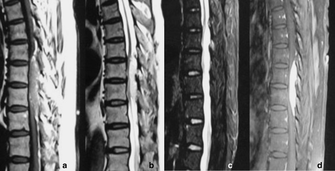
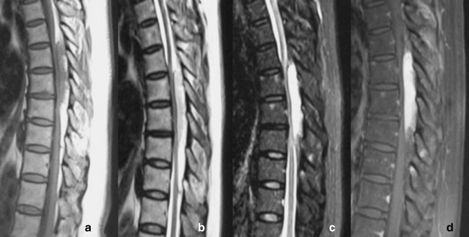
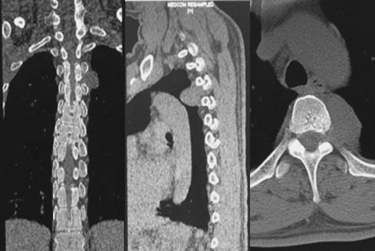
Results: Twenty-one patients were described. We divide SAs into two types: "intraspinal" and "dumbbell-shaped". The former were further subclassified as "with lipomatosis" and "without lipomatosis". Overweight people are more likely to get the "with lipomatosis" type which needs different surgical strategy and/or a diet therapy to get better outcomes.
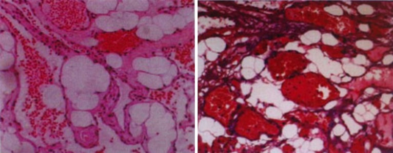
Conclusion: Diagnosis of SAs should be made with reference to clinical, radiological, and pathological findings. Application of different methods is needed to treat SAs.
Figures
References
-
- Kujas M, Lopes M, Lalam TF, et al. Infiltrating extradural spinal angiolipoma. Clin Neuropathol. 1999;18:93–98. - PubMed
-
- Fourney DR, Tong KA, Macaulay RJB, et al. Spinal angiolipoma. Can J Neu Sci. 2001;28:82–88. - PubMed
-
- Ando K, Imagama S, Wakao N, et al. Examination of the influence of ossification of the anterior longitudinal ligament on symptom progression and surgical outcome of ossification of the thoracic ligamentum flavum: a multicenter study. J Neurosurg Spine. 2012;16:147–153. doi: 10.3171/2011.10.SPINE11296. - DOI - PubMed
-
- Association JO Scoring system for cervical myelopathy. J Jpn Orthop Assoc. 1994;68:490–503.
MeSH terms
LinkOut - more resources
Full Text Sources
Other Literature Sources
