. 2013 Nov 18;210(12):2569-82.
doi: 10.1084/jem.20131241. Epub 2013 Nov 4.

A molecular basis for the association of the HLA-DRB1 locus, citrullination, and rheumatoid arthritis

Affiliations

A molecular basis for the association of the HLA-DRB1 locus, citrullination, and rheumatoid arthritis

Stephen W Scally et al. J Exp Med. .

Abstract

Rheumatoid arthritis (RA) is strongly associated with the human leukocyte antigen (HLA)-DRB1 locus that possesses the shared susceptibility epitope (SE) and the citrullination of self-antigens. We show how citrullinated aggrecan and vimentin epitopes bind to HLA-DRB1*04:01/04. Citrulline was accommodated within the electropositive P4 pocket of HLA-DRB1*04:01/04, whereas the electronegative P4 pocket of the RA-resistant HLA-DRB1*04:02 allomorph interacted with arginine or citrulline-containing epitopes. Peptide elution studies revealed P4 arginine-containing peptides from HLA-DRB1*04:02, but not from HLA-DRB1*04:01/04. Citrullination altered protease susceptibility of vimentin, thereby generating self-epitopes that are presented to T cells in HLA-DRB1*04:01(+) individuals. Using HLA-II tetramers, we observed citrullinated vimentin- and aggrecan-specific CD4(+) T cells in the peripheral blood of HLA-DRB1*04:01(+) RA-affected and healthy individuals. In RA patients, autoreactive T cell numbers correlated with disease activity and were deficient in regulatory T cells relative to healthy individuals. These findings reshape our understanding of the association between citrullination, the HLA-DRB1 locus, and T cell autoreactivity in RA.


Figures
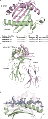
Figure 1.
Figure 1.
HLA-DRB1*04:01 in complex with Vimentin-71Cit66-78. (a) Polymorphic residues involved in susceptibility to RA. The peptide-binding groove of an HLA-DR molecule is shown in cartoon representation with the α-chain colored in green and the β-chain colored in pink. Residues Val11β, His13β, Lys71β, and Ala74β are represented as sticks and correspond to the residues present in HLA-DR401, the HLA with the highest risk associated with RA. (b) Sequence alignment of the three HLA-DRB1*04 alleles used in this study showing amino acid polymorphisms. “-” indicates residue conserved with that of HLA-DRB1*04:01:01. Val11β, His13β are conserved in all three alleles (not depicted). (c) HLA-DRB1*04:01 in complex with vimentin-71Cit66-78. The vimentin-71Cit66-78 peptide is bound in the peptide-binding groove, with carbons colored in yellow, nitrogens colored in blue, and oxygens colored in red. The α and β chains are shown in cartoon representation, and colored in green and pink, respectively. (d) Side view of the bound vimentin-71Cit66-78 peptide. The peptide’s 2Fo-Fc electron density map is shown in blue and contoured to 1 σ, showing unambiguous density for the peptide. Peptide residues are labeled and numbered, with Citrulline71 occupying the P4 pocket.
Figure 2.
Figure 2.
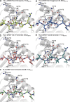
Side view of epitopes bound to HLA-DR4. (a) HLA-DRB1*04:01 bound to vimentin-64Cit59-71. (b) HLA-DRB1*04:01 bound to vimentin-64-69-71Cit59-71. (c) HLA-DRB1*04:01 bound to aggrecan-93-95Cit89-103. (d) HLA-DRB1*04:04 bound to vimentin-71Cit59-71. (e) HLA-DRB1*04:02 bound to vimentin-71Cit66-78. (f) HLA-DRB1*04:02 bound to Vimentin66-78. The peptide’s 2Fo-Fc electron density map is shown in blue and contoured to 1 σ. Peptide residues are labeled and numbered.
Figure 3.
Figure 3.
Interactions with citrulline in the P4 pocket of HLA-DRB1*04:01 and HLA-DRB1*04:04. (a) Vimentin-71Cit66-78 colored in yellow, (b) vimentin-64Cit59-71 colored in pink, (c) vimentin-64-69-71Cit59-71 colored in green, and (d) aggrecan-93-95Cit89-103 (colored in blue, bound to HLA-DRB1 *04:01). Residues from the β chain important for contacts with the P4 citrulline are represented as sticks. (e) Vimentin-71Cit66-78 colored in teal bound to HLA-DRB1*04:04.
Figure 4.
Figure 4.
Comparison of the interactions between citrulline and arginine in the P4 pocket of HLA-DRB1*04:02. (a) Vimentin-71Cit66-78 colored in green; (b) Vimentin66-78 colored in purple. The solvent-accessible electrostatic potential was calculated for panel c HLA-DRB1*04:01 and (d) HLA-DRB1*04:02 bound to vimentin-71Cit66-78. Electrostatic calculations were performed using APBS (±12 kT/e).
Figure 5.
Figure 5.
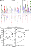
HLA-DRB1*04 binding motifs and protease sensitivity of citrullinated epitopes. (a) HLA Binding motifs of DRB1*04:01, DRB1*04:02 and DRB1*04:04 were generated from immunoaffinity purified allotypes isolated from T2-DRB1*04:01, 04:02 and 04:04 cells expressing DM. Each HLA DR allotype was affinity purified, and bound peptides were isolated and analyzed by Liquid chromatography–mass spectrometry (LC-MS/MS). To generate peptide-binding motifs, the minimal core sequences found within nested sets were extracted and the resulting list of peptides aligned and visualized using Icelogo. Positively associated residues (P > 0.05) at each relative position are shown above the x-axis and negatively associated residues are shown below. Residues height is proportionate to prevalence, with residues shown in pink having infinite height reflecting absolute presence or absence at that position in the bound peptides. (b) Citrullination alters cleavage of vimentin by Cathepsin L. Recombinant human vimentin was citrullinated in vitro, and the Cathepsin L digestion patterns of native and citrullinated vimentin were observed by LC-MS/MS. Observed cleavages are highlighted by arrows in the region of vimentin-spanning residues 51–81. The amount of selected peptides (as determined by area under the curve quantitation for extracted ion chromatograms) from this region that span the immunogenic 59–71 region of vimentin are shown as a function of digestion time (1, 5, 3, and 60 min digests).
Figure 6.
Figure 6.
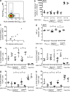
CD4+tetramer+ T cells circulate in RA patients and healthy controls. (a) PBMC from a representative HLA-DRB1*04:01+ RA patient were stained with PE-labeled HLA-DRB1*04:01-HA306-318 tetramer and Brilliant Violet 421 (BV)-labeled HLA-DRB1*04:01-vimentin-64Cit59-71 tetramer and FITC-labeled anti-CD11c, CD14, CD16, and CD19 and APC/Cy7-labeled anti-CD4, and then analyzed by flow cytometry, setting gates on FITCCD4+ cells based on PE- and BV-fluorescence minus one (FMO) staining. PBMCs from 9 HLA-DRB1*04:01+ RA patients (RA, filled circles) and 4 healthy controls (HC, empty circles) were stained with AQUA live dead discriminator, PE-labeled HLA-DRB1*04:01-HA306-318, -vimentin-64Cit59-71, or -aggrecan-93-95Cit89-103 tetramers, Alexa Fluor 488 Foxp3, PerCP/Cy5.5-CD14, Pacific blue-CD45RO, APC-CD28, APC/Cy7-CD4, PE/Cy7-CD25, and count beads were added. Either CD4+CD14tetramer+ or total CD4+ T cells were gated and the frequency of CD4+CD14tetramer+ or total CD4+CD14 cells/ml blood was calculated (b). The number of HLA-DRB1*04:01-vimentin-64Cit59-71–specific T cells was plotted relative to the four variable disease activity score (DAS4vCRP) of each RA patient (c). MFI of tetramer+ T cells (d), percentage of CD4+CD45ROCD25+Foxp3+ resting T reg cells (e), CD4+CD45RO+CD25hiFoxp3+ activated T reg cells (f), CD4+CD45ROCD25Foxp3 naive T cells (g) or CD4+CD45RO+CD25Foxp3 effector/memory T cells (h) of RA patients (columns 1, 3, 5, and 7) and healthy controls (columns 2, 4, 6, and 8) is shown for each or tetramer+ population (columns 1–6) and for total PB CD4+ T cells (columns 7 and 8). *, P < 0.05; **, P < 0.01, using the Mann-Whitney test to compare RA patients and healthy controls. The number of PB HLA-DRB1*04:01-vimentin-64Cit59-71–specific T cells were correlated with disease activity score in RA patients (Spearman r = 0.76; P < 0.05).

References

    1. Abadie V., Sollid L.M., Barreiro L.B., Jabri B. 2011. Integration of genetic and immunological insights into a model of celiac disease pathogenesis. Annu. Rev. Immunol. 29:493–525 10.1146/annurev-immunol-040210-092915 - DOI - PubMed
    1. Aletaha D., Neogi T., Silman A.J., Funovits J., Felson D.T., Bingham C.O., III, Birnbaum N.S., Burmester G.R., Bykerk V.P., Cohen M.D., et al. 2010. Rheumatoid arthritis classification criteria: an American College of Rheumatology/European League Against Rheumatism collaborative initiative. Arthritis Rheum. 62:2569–2581 10.1002/art.27584 - DOI - PubMed
    1. Aricescu A.R., Lu W., Jones E.Y. 2006. A time- and cost-efficient system for high-level protein production in mammalian cells. Acta Crystallogr. D Biol. Crystallogr. 62:1243–1250 10.1107/S0907444906029799 - DOI - PubMed
    1. Bailey T.L., Boden M., Buske F.A., Frith M., Grant C.E., Clementi L., Ren J., Li W.W., Noble W.S. 2009. MEME SUITE: tools for motif discovery and searching. Nucleic Acids Res. 37:W202–8 10.1093/nar/gkp091 - DOI - PMC - PubMed
    1. Bharadwaj M., Illing P., Theodossis A., Purcell A.W., Rossjohn J., McCluskey J. 2012. Drug hypersensitivity and human leukocyte antigens of the major histocompatibility complex. Annu. Rev. Pharmacol. Toxicol. 52:401–431 10.1146/annurev-pharmtox-010611-134701 - DOI - PubMed

Publication types

MeSH terms