. 1986 Mar;63(3):274-88.
doi: 10.1016/0013-4694(86)90095-7.

Behavioral states in the chronic medullary and midpontine cat

Behavioral states in the chronic medullary and midpontine cat

J M Siegel et al. Electroencephalogr Clin Neurophysiol. 1986 Mar.

Abstract

Behavioral state organization was studied in the caudal portion of chronically maintained cats with transections at the ponto-medullary junction or midpontine level. The cats spent most of their time in a 'quiescent state.' This state was periodically interrupted by 'phasic activations.' During quiescence, ECG and reticular unit activity rates were low and regular. EMG levels resembled those seen during non-REM sleep in intact cats. During phasic activations, unit activity in the nucleus gigantocellularis and neck EMG activity increased to levels seen in the intact cat during active waking. Gross postural changes, vestibular slow phase head nystagmus and head shake reflexes could be observed at these times. No periods of neck muscle atonia were observed in either state. No periods of brain-stem controlled rapid eye movements (REMs) occurred. Unit activity patterns similar to those seen in the intact cat during REM sleep were never observed. Physostigmine administration did not produce REM sleep signs, but rather, triggered an aroused state. Phasic activations occurred in a regular ultradian rhythm, with a period similar to that seen in the REM sleep cycle. We conclude that the chronic medullary cat retains primitive aroused and quiescent states, but does not have any of the local signs of REM sleep. However, the medulla does have the capability of generating ultradian rhythmicities which may contribute to the control of the basic rest activity cycle and the REM, non-REM sleep cycle.


Figures

Fig. 1.
Fig. 1.
Levels of transection of all cats employed in the study. RN, red nucleus; LC, locus coeruleus; 7, genu of facial nerve; 6, abducens nucleus; IO, inferior olive. Stereotaxic coordinates derived from sagittal plates of Berman (1968) atlas at L1.2.
Fig. 2.
Fig. 2.
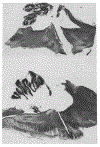
Top: sagittal section at 1.2 mm lateral to midline through the transection in cat 24. Abducens nucleus is prominent caudal to transection. Cresyl violet stain. Bottom: sagittal section at 1.2 mm lateral to midline through the transection in cat 25.
Fig. 3.
Fig. 3.
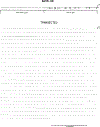
States seen in the chronic medullary cat. EMG, dorsal neck electromyogram; EKG, electrocardiogram; Resp, thoracic strain gauge. Samples were taken from day 7 in cat 27. EMG calibration, 50 μV.
Fig. 4.
Fig. 4.
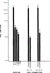
Nuchal EMG amplitude in intact cats during active waking (A-WA), quiet waking (Q-WA), light and deep slow wave sleep (SWS-1, SWS-2) and REM sleep, compared to EMG levels seen during arousals and in quiescent state after transection. Amplitude levels based on 10 samples taken in each state in both baseline and post-transection recordings in cats 25, 27, 31 and 46.
Fig. 5.
Fig. 5.
Abducens controlled eye movements prior to and after brain-stem transection. In baseline recording, periods of rapid eye movement can be observed during REM sleep. After transection only isolated nictitating membrane blinks are observed. Calibration, 100 μV.
Fig. 6.
Fig. 6.
Locations of medullary units recorded after transection. Squares, units with rates > 4.0/sec in quiescence; circles, units with rates < 4.0/sec. Empty square, unit decreasing rate during phasic arousal.
Fig. 7.
Fig. 7.
Compressed display of medullary reticular formation (RF) unit discharge rate during sleep-waking cycle in intact cat, and after brain-stem transection. Tracing is output of a digital counter indicating number of action potentials and resetting at 1 sec intervals. Note the long periods of accelerated and irregular unit discharge rate during waking and REM sleep in the intact cat. RF cells in the transected cats have extremely regular discharge rates with 1 sec counts virtually identical for long time periods. Short periods of increased unit activity occur in conjunction with phasic arousals.
Fig. 8.
Fig. 8.
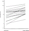
Increases in discharge rates of medullary units with phasic arousal from quiescent state baseline. Rate is in spikes/sec. Unit activity rates are based on 10 consecutive 10 sec intervals during quiescence and on the mean of 1 sec samples during 2 phasic arousals.
Fig. 9.
Fig. 9.
Periodicities in intact control and in transected animals. The left column of the figure presents spectral power density plots of REM sleep onset periodicities in intact control animals. The right columns of the figure present spectral density plots of phasic arousals in 2 representative animals taken 1, 2 and 3 weeks after transection. In control animals, the spectral power density plot has its peaks between 30 and 60 min. One week after transection, no prominent peak is visible in the spectral power density of either animal and the equivalent noise bandwidth (ENB) is correspondingly high. By the third week, prominent peaks are present at between 30 and 60 rain in both transected animals. ENB values are reduced and are comparable to values in intact animals.

Similar articles

Cited by

References

    1. Berman AL, The Brain Stem of the Cat. University of Wisconsin Press, Madison, WI, 1968.
    1. Bonvallet M and Bloch V Bulbar control of cortical arousal. Science, 1976, 133: 1133–1134. - PubMed
    1. De Andres I and Reinoso-Suarez F Participation of the cerebellum in the regulation of the sleep-wakefulness cycle through the superior cerebellum peduncle. Arch. ital. Biol, 1979, 117: 140–163. - PubMed
    1. Delorme F, Vimont P, et Jouvet D. Etude statistique du cycles veille-sommeils chez le chat. C.R. Soc. Biol. (Paris), 1964, 158: 2128–2130. - PubMed
    1. Dixon WJ, Brown MB, Engelman L, Frane JW, Hill MA, Jennrich RI and Toporek JD BMDP Statistical Software. University of California Press, Berkeley, CA, 1983.

Publication types

LinkOut - more resources