Review
. 2013 Nov;195(3):643-81.
doi: 10.1534/genetics.113.153197.

Ribosome biogenesis in the yeast Saccharomyces cerevisiae

Affiliations
Review

Ribosome biogenesis in the yeast Saccharomyces cerevisiae

John L Woolford Jr et al. Genetics. 2013 Nov.

Abstract

Ribosomes are highly conserved ribonucleoprotein nanomachines that translate information in the genome to create the proteome in all cells. In yeast these complex particles contain four RNAs (>5400 nucleotides) and 79 different proteins. During the past 25 years, studies in yeast have led the way to understanding how these molecules are assembled into ribosomes in vivo. Assembly begins with transcription of ribosomal RNA in the nucleolus, where the RNA then undergoes complex pathways of folding, coupled with nucleotide modification, removal of spacer sequences, and binding to ribosomal proteins. More than 200 assembly factors and 76 small nucleolar RNAs transiently associate with assembling ribosomes, to enable their accurate and efficient construction. Following export of preribosomes from the nucleus to the cytoplasm, they undergo final stages of maturation before entering the pool of functioning ribosomes. Elaborate mechanisms exist to monitor the formation of correct structural and functional neighborhoods within ribosomes and to destroy preribosomes that fail to assemble properly. Studies of yeast ribosome biogenesis provide useful models for ribosomopathies, diseases in humans that result from failure to properly assemble ribosomes.

Keywords: RNA polymerase I; ribosomal RNA; ribosome; ribosome assembly; snoRNAs.


Figures

Figure 1
Figure 1
(A) Secondary structure of S. cerevisiae 18S, 25S, and 5.8S rRNAs. Left, the four domains of 18S rRNA secondary structure are indicated in different colors. Right, 25S rRNA contains six domains (I–VI) of secondary structure. The 5.8S rRNA (black) base pairs with domain I (adapted from www.rna.ccbb.utexas.edu). These secondary structures are phylogenetically conserved throughout all kingdoms, although eukaryotic rRNAs contain expansion segments not found in prokaryotic or archaeal rRNAs. (B) Tertiary structures of 18S rRNA (left) and 25S plus 5.8S rRNAs (right), from the crystal structure of yeast ribosomes (Ben-Shem et al. 2011).
Figure 2
Figure 2
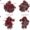
Crystal structure at 3.0-A resolution of S. cerevisiae 40S and 60S ribosomal subunits. (A) Views of the solvent-exposed surface (left) and subunit interface (right) of the 60S subunit. CP, central protuberance. (B) The solvent-exposed surface (left) and the subunit interface (right) of the 40S subunit. rRNA is represented in gray and r-proteins are in red. The crystal structure is adapted from Protein Data Bank files 3U5B, 3U5C, 3U5D, and 3U5E from Ben-Shem et al. (2011).
Figure 3
Figure 3
Organization of the rDNA locus in S. cerevisiae. The rDNA repeats (150–200) are located on chromosome 12. A single repeated unit is transcribed by RNA polymerase I (RNA pol I) to synthesize the 35S primary pre-rRNA transcript that will be processed to produce the mature 18S, 5.8S, and 25S rRNAs (arrow pointing right) and by RNA polymerase III (RNA pol III) to synthesize the 5S rRNA (arrow pointing left). NTS, nontranscribed spacer; ETS, external transcribed spacer; ITS, internal transcribed spacer.
Figure 4
Figure 4
Yeast chromatin spreads of nucleolar contents analyzed by electron microscopy. Transcription of the repeated rDNA units can be visualized as “Christmas trees”, where the trunk of the tree is the rDNA, the branches are the rRNA, and the ornaments are the knobs on the 5′ ends. The knob contains the SSU processome. Yeast chromatin spreads are courtesy of Sarah French and Ann Beyer.
Figure 5
Figure 5
Pathway for processing of yeast pre-rRNA. The 35S pre-rRNA is transcribed by RNA polymerase I and contains sequences for 18S, 5.8S, and 25S rRNAs (black, dark gray, and light gray cylinders) flanked and separated by external and internal transcribed spacers (ETS and ITS, solid lines). Pre-5S rRNA (white cylinder) is transcribed by RNA polymerase III. Spacer sequences are removed from pre-rRNA by the indicated series of endonucleolytic and exonucleolytic processing steps, within assembling ribosomes. Each processing site is indicated. Processing begins in the nucleolus of the cell, but later steps occur in the nucleoplasm and cytoplasm. Note that in rapidly dividing cells, the majority of pre-rRNA undergoes cotranscriptional cleavage at the A0, A1, and A2 sites before transcription is completed.
Figure 6
Figure 6
Box H/ACA and box C/D snoRNAs target pre-RNA modification via base pairing. A Box H/ACA snoRNA is shown on the left. A box C/D snoRNA is shown on the right. snoRNAs are indicated in black. Target rRNAs are indicated in red. This diagram is adapted from Figure 1 of Watkins and Bohnsack (2012).
Figure 7
Figure 7
Conformational switch of pre-rRNA involving the removal of ITS1 spacer RNA. Left, sequences in the 3′ end of ITS1 spacer (gray) are predicted to base pair with nucleotides in what becomes the 5′ end of 5.8S rRNA (black). Right, in mature 60S subunits, these same sequences in 5.8S rRNA base pair with nucleotides in 25S rRNA to form helix 2, to which r-protein L17 binds. Removal of the 3′ end of ITS1 from 27SA3 pre-rRNA may be required to form a stable neighborhood including helix 2. Alternatively, remodeling of preribosomes to form a stable helix 2, including bound L17, may enable 5′ exonucleolytic processing of 27SA3 pre-rRNA.
Figure 8
Figure 8
Pathway for maturation of preribosomes to form 40S and 60S ribosomal subunits. Sequential assembly intermediates are shown, distinguished by the pre-rRNA processing intermediates contained within them. Most r-proteins (light blue) and many assembly factors (dark blue) associate with the early nucleolar/nuclear precursor particles. Some assembly factors join preribosomes in middle steps of assembly or even during late steps in the cytoplasm. Release of assembly factors from preribosomes occurs at early, middle, or late stages of subunit maturation.
Figure 9
Figure 9
Hierarchical pathway for stable association with preribosomes of assembly factors and some r-proteins necessary for early and middle steps of 60S subunit assembly. Proteins that form the Pwp1 subcomplex (blue) function together to process 27SA2 and 27SA3 pre-rRNAs and are required for stable association of A3 factors (green). The A3 factors are interdependent for their association with preribosomes and are necessary for 27SA3 pre-rRNA processing. The presence of the A3 factors is required for assembly of the DEAD-box proteins Drs1 and Has1 (light blue). In turn, these two factors are necessary for the stable association with preribosomes of r-proteins L17, L26, L35, and L37 (purple), which bind to helices formed between 5.8S and 25S rRNAs. Both the A3 factors and r-proteins bound to the 5′ end of 5.8S rRNA are required for the stable association with preribosomes of a subset of factors required for 27SB pre-rRNA processing (red). The B factors assemble into preribosomes in two parallel pathways that converge to recruit the GTPase Nog2.
Figure 10
Figure 10
Locations on late pre-60S ribosomes of export factors Mex67/Mtr2, Nmd3, and Arx1 and assembly factors Tif6 and Rei1. Cryo-EM reconstructions of particles purified using TAP-tagged Arx1 were modeled onto the crystal structure of the mature 60S ribosomal subunit. This model is adapted from Figure 2 of Bradatsch et al. (2012).
Figure 11
Figure 11
Pathway for late steps of maturation of pre-60S subunits in the cytoplasm. Assembly factors Nog1, Rlp24, Arx1, Alb1, Mrt4, Tif6, and Nmd3 are sequentially released from 66S preribosomes in the cytoplasm by the ATPase Drg1; and Ssa/Ssb by the GTPases Efl1 and Lsg1; and the phosphatase Yvh1 with the aid of Jjj1, Rei1, and Sdo1. This pathway also enables late assembly of r-proteins L24 and P0, replacing their homologues Rlp24 and Mrt4, respectively. This pathway is adapted from Figure 7 of Lo et al. (2010).
Figure 12
Figure 12
Assembly factors prevent premature association of translation initiation factors with pre-40S ribosomes. Left, Tsr1, Rio2, and Dim1 are located on the subunit interface of pre-40S ribosomes, overlapping with the binding sites of translation initiation factors eIF1 (blue) and eIF1A (binding site in red) and the P-site tRNA (green). Center, Nob1 and Pno1/Dim2 bound to pre-40S ribosomes prevent association of eIF3 (purple). Right, The presence of Ltv1 and Enp1 on the solvent side of pre-40S particles prevents proper interaction of r-protein S3 with helix 16, thus blocking opening of the mRNA entry channel. This diagram is adapted from Figure 1 of Karbstein (2013).

Similar articles

Cited by

References

    1. Adams C. C., Jakovljevic J., Roman J., Harnpicharnchai P., Woolford J. L., Jr, 2002. Saccharomyces cerevisiae nucleolar protein Nop7p is necessary for biogenesis of 60S ribosomal subunits. RNA 8: 150–165 - PMC - PubMed
    1. Allmang C., Tollervey D., 1998. The role of the 3′ external transcribed spacer in yeast pre- rRNA processing. J. Mol. Biol. 278: 67–78 - PubMed
    1. Allmang C., Kufel J., Chanfreau G., Mitchell P., Petfalski E., et al. , 1999. Functions of the exosome in rRNA, snoRNA and snRNA synthesis. EMBO J. 18: 5399–5410 - PMC - PubMed
    1. Allmang C., Mitchell P., Petfalski E., Tollervey D., 2000. Degradation of ribosomal RNA precursors by the exosome. Nucleic Acids Res. 28: 1684–1691 - PMC - PubMed
    1. Armache J. P., Jarasch A., Anger A. M., Villa E., Becker T., et al. , 2010a Cryo-EM structure and rRNA model of a translating eukaryotic 80S ribosome at 5.5-A resolution. Proc. Natl. Acad. Sci. USA 107: 19748–19753 - PMC - PubMed

Publication types

MeSH terms

LinkOut - more resources