. 2014 Jan;34(1):132-45.
doi: 10.1128/MCB.00639-13. Epub 2013 Nov 4.

A role for USP7 in DNA replication

Affiliations

A role for USP7 in DNA replication

Madhav Jagannathan et al. Mol Cell Biol. 2014 Jan.

Abstract

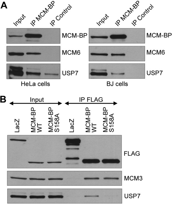
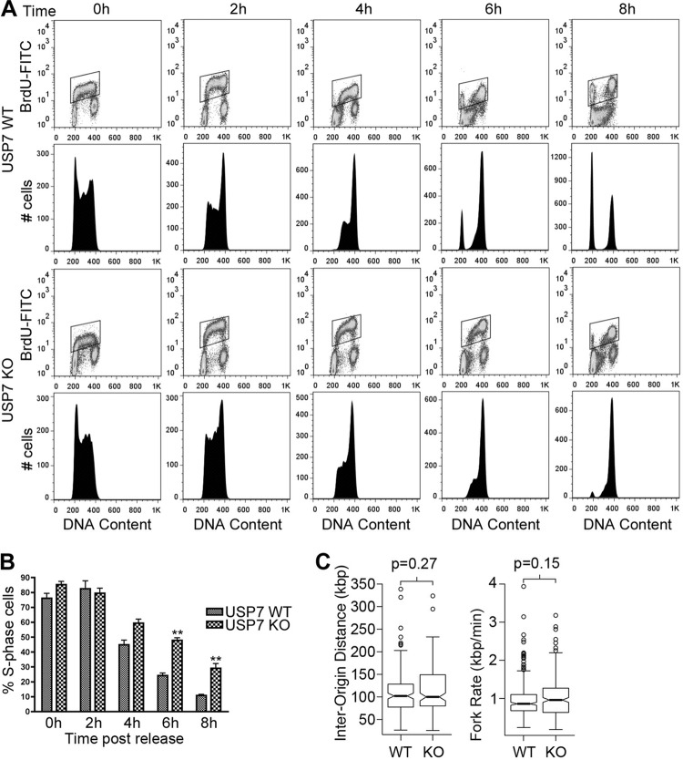
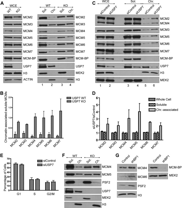
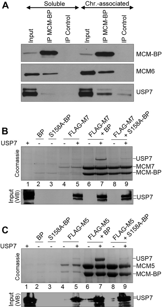
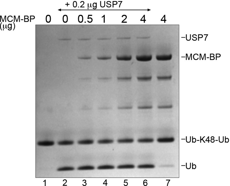
The minichromosome maintenance (MCM) complex, which plays multiple important roles in DNA replication, is loaded onto chromatin following mitosis, remains on chromatin until the completion of DNA synthesis, and then is unloaded by a poorly defined mechanism that involves the MCM binding protein (MCM-BP). Here we show that MCM-BP directly interacts with the ubiquitin-specific protease USP7, that this interaction occurs predominantly on chromatin, and that MCM-BP can tether USP7 to MCM proteins. Detailed biochemical and structure analyses of the USP7-MCM-BP interaction showed that the (155)PSTS(158) MCM-BP sequence mediates critical interactions with the TRAF domain binding pocket of USP7. Analysis of the effects of USP7 knockout on DNA replication revealed that lack of USP7 results in slowed progression through late S phase without globally affecting the fork rate or origin usage. Lack of USP7 also resulted in increased levels of MCM proteins on chromatin, and investigation of the cause of this increase revealed a defect in the dissociation of MCM proteins from chromatin in mid- to late S phase. This role of USP7 mirrors the previously described role for MCM-BP in MCM complex unloading and suggests that USP7 works with MCM-BP to unload MCM complexes from chromatin at the end of S phase.


Figures

FIG 1
FIG 1
Coimmunoprecipitation of USP7 with MCM-BP. (A) Equal amounts of whole-cell extracts from HeLa cells or BJ cells were incubated with antibody against MCM-BP or nonspecific IgG (control). Immunoprecipitants were analyzed by Western blotting against the indicated proteins. Note that USP7 is known to run as a doublet due to the presence of two isoforms (69). (B) HeLa cells were transfected with FLAG-LacZ, FLAG–MCM-BP, and FLAG–MCM-BP S158A expression plasmids and 48 h later were subjected to anti-FLAG immunoprecipitation. Western blots for the indicated proteins are shown for the immunoprecipitates (IP FLAG) and the starting cell lysates (5% input).
FIG 2
FIG 2
Gel filtration analyses of USP7–MCM-BP complexes. (A) Schematic representation of the domain organization of USP7, showing amino acid numbers. (B to E) Purified MCM-BP was incubated with equimolar quantities of purified full-length USP7 (B), USP7 1–540 (C), USP7 1–205 (D), or USP7 202–540 (E). In panel F, purified MCM-BP S158A was incubated with an equimolar quantity of purified full-length USP7. All protein mixtures were run through a Suparose gel filtration column, and equal-volume fractions were collected. Equal volumes of each of the indicated fractions were then analyzed by SDS-PAGE and Coomassie staining. The positions of the gel filtration standards myoglobin (17 kDa), ovalbumin (44 kDa), aldolase (158 kDa), and thyroglobulin (669 kDa) on the same column are indicated at the top of each panel.
FIG 3
FIG 3
GST pulldown assays of USP7–MCM-BP interactions. Purified MCM-BP was incubated with equimolar quantities of purified GST (A), GST-USP7 1–205 (B), GST-USP7 202–560 (C), GST-USP7 560–1102 (D), GST-USP7 1–540 (E), GST-USP7 1–560 (F), or GST-USP7 1–560 DW (G) that was immobilized on glutathione-agarose beads. In panel H, MCM-BP S158A was incubated with an equimolar quantity of purified GST USP7 1–560. Input and 1/10 flowthrough for GST-tagged proteins are indicated by L1 and F1, respectively. Input and 1/10 flowthrough for MCM-BP/MCM-BP S158A are indicated by L2 and F2, respectively. Bound proteins were eluted with two glutathione incubations (E1 and E2) and analyzed by SDS-PAGE and Coomassie staining.
FIG 4
FIG 4
Crystal structure of USP7 54–205 bound to MCM-BP peptide 152RVSPSTSYTP161. (A) Ribbon representation of USP7 54–205 (cyan) bound to the MCM-BP peptide, shown in stick representation (yellow). PDB ID 4KG9. (B) Detailed interactions of MCM-BP (yellow), with β-strand 7 of USP7 54–205 (cyan) shown in stick representation. The hydrogen bonds are shown with black dashed lines. (C) Overlay of MCM-BP peptide (yellow) with Mdm2 396QPSTSS401 peptide (green) from previously determined Mdm2-USP7 structure (38).
FIG 5
FIG 5
USP7 affects progression through late S phase. (A) WT and USP7-null cells were pulsed with BrdU and chased in the absence of BrdU for the indicated times. The DNA content of BrdU-positive cells was measured by flow cytometry at each time point. For each sample, the polygon in the top panel shows BrdU-incorporated cells, while the bottom panel shows cell phase distribution as determined by DNA content. (B) The percentages of S phase cells from three independent experiments performed as for panel A were determined using FlowJo software (Treestar Inc.) and graphed. Error bars represent standard deviations, and “∗∗” represents P < 0.01. (C) Distributions of interorigin distance (IOD) and replication fork progression were obtained by DNA combing in USP7-null (KO) and WT HCT116 cells. Median values for IOD are 102 kbp (WT) and 100 kbp (KO). Median values for the fork rate are 0.86 kbp/min (WT) and 0.96 kbp/min (KO). P values were determined by a two-tailed Mann-Whitney U test used to compare nonnormal distributions.
FIG 6
FIG 6
USP7 depletion increases the chromatin association of MCM proteins. (A) Whole-cell (WCE), soluble (Sol.), and chromatin-associated (Chr.) lysates from WT and USP7-null (KO) cells were analyzed by SDS-PAGE and Western blotting against the indicated MCM proteins and USP7. In the left panel, histone H3 and actin are used as loading controls. (B) MCM protein bands from the soluble and chromatin-associated lysate were quantified from three independent experiments by densitometry (GelEval software). The ratio of chromatin-associated to soluble protein was calculated for each MCM protein from the WT and USP7-null cells and graphed. Error bars represent standard deviations. (C) Whole-cell, soluble, and chromatin-associated lysates from HeLa cells transfected with control or USP7-targeted siRNA (siControl and siUSP7, respectively) were analyzed by SDS-PAGE and Western blotting for the indicated MCM proteins and USP7. Blots against H3 (chromatin associated) and MEK2 (soluble) are used to indicate appropriate fractionation and also serve as loading controls. Note that since only a small proportion of MCM-BP is chromatin associated, the amount of chromatin lysate loaded in this experiment is not enough to detect MCM-BP in this fraction. (D) MCM protein bands from the whole-cell, soluble, and chromatin-associated lysate were quantified from two independent experiments by densitometry. The ratio of siUSP7 band density to siControl band density was calculated for each MCM protein in the whole-cell, soluble, and chromatin-associated lysate and graphed. Error bars represent standard deviations. (E) DNA content of HeLa cells transfected with control or USP7-targeted siRNA was measured by flow cytometry in three independent experiments and graphed using FlowJo software (Treestar Inc.). The percentages of cells in G1, S, and G2/M were estimated using FlowJo software and are shown in the graph. Error bars represent standard deviations. (F) Soluble (Sol.) and chromatin-associated (Ch.) lysates from WT and USP7-null cells (KO) were analyzed by SDS-PAGE and Western blotting against the indicated MCM proteins, Psf2 and USP7. Histone H3 (chromatin-associated protein) and MEK2 (soluble protein) indicate appropriate fractionation and also serve as loading controls. (G) Chromatin-associated lysates from HeLa cells transfected with control plasmid or shMCM-BP1 were analyzed by SDS-PAGE and Western blotting for the indicated MCM proteins, Psf2, and histone H3 as a loading control (left panel). Since only a small proportion of MCM-BP is chromatin associated, MCM-BP silencing was assessed by comparing equal volumes of the soluble lysate, compared to MEK2 as a loading control (right panel).
FIG 7
FIG 7
USP7 depletion affects unloading of the MCM complex at the end of S phase. (A) HeLa cells transfected with control or USP7-targeted siRNA were synchronized to the G1/S boundary using a double thymidine block and released for the indicated times (0 h to 8 h). The chromatin-associated extract for each time point was analyzed by SDS-PAGE and Western blotting for the indicated MCM proteins and USP7. PCNA was used as a loading control. (B) The DNA content of the above samples was measured using flow cytometry and graphed using FlowJo software (Treestar Inc.). (C) MCM5 protein bands from the chromatin-associated fraction at 0 and 8 h after release from a double thymidine block were quantified from three independent experiments by densitometry. Average values with standard deviations are shown relative to results for 0-h siControl samples (set to 100). ∗, P < 0.05.
FIG 8
FIG 8
MCM-BP binds USP7 on chromatin and can mediate an interaction between the USP7 and MCM proteins. (A) Immunoprecipitations were performed with soluble and chromatin-associated lysates from HeLa cells using antibody against MCM-BP or negative-control IgG. Immunoprecipitants were analyzed by SDS-PAGE and Western blotting against the indicated proteins. (B and C) FLAG-tagged MCM7 (B) or FLAG-tagged MCM5 (C) was expressed individually or coexpressed with MCM-BP (BP) or MCM-BP S158A (S158A-BP) in insect cells. In each case, these proteins were expressed with and without USP7, as indicated. Insect cell lysates were then incubated with anti-FLAG resin to recover FLAG-MCM7 or FLAG-MCM5 with associated proteins. Bound proteins were eluted with protein sample buffer and analyzed by SDS-PAGE and Coomassie staining (top panel) or Western blotting for USP7 (bottom panel).
FIG 9
FIG 9
MCM-BP does not inhibit the catalytic activity of USP7. Purified USP7 (0.2 μg) was preincubated with increasing amounts of MCM-BP (as indicated) and then incubated with K48-linked diubiquitin (3 μg) for 10 min at 37°C. A Coomassie-stained SDS-PAGE gel is shown, where the positions of the diubiquitin substrate (Ub-K48-Ub) and monoubiquitin product (Ub) are indicated. Lanes 1 and 7 are controls lacking USP7.

Similar articles

Cited by

References

    1. Boos D, Frigola J, Diffley JF. 2012. Activation of the replicative DNA helicase: breaking up is hard to do. Curr. Opin. Cell Biol. 24:423–430. 10.1016/j.ceb.2012.01.011 - DOI - PubMed
    1. Burkhart R, Schulte D, Hu D, Musahl C, Gohring F, Knippers R. 1995. Interactions of human nuclear proteins P1Mcm3 and P1Cdc46. Eur. J. Biochem. 228:431–438. 10.1111/j.1432-1033.1995.tb20281.x - DOI - PubMed
    1. Lei M, Kawasaki Y, Tye BK. 1996. Physical interactions among Mcm proteins and effects of Mcm dosage on DNA replication in Saccharomyces cerevisiae. Mol. Cell. Biol. 16:5081–5090 - PMC - PubMed
    1. Rowles A, Chong JP, Brown L, Howell M, Evan GI, Blow JJ. 1996. Interaction between the origin recognition complex and the replication licensing system in Xenopus. Cell 87:287–296. 10.1016/S0092-8674(00)81346-X - DOI - PubMed
    1. Mahbubani HM, Chong JP, Chevalier S, Thommes P, Blow JJ. 1997. Cell cycle regulation of the replication licensing system: involvement of a Cdk-dependent inhibitor. J. Cell Biol. 136:125–135. 10.1083/jcb.136.1.125 - DOI - PMC - PubMed

Publication types

MeSH terms

LinkOut - more resources