. 2014 Jan;34(2):196-209.
doi: 10.1128/MCB.00664-13. Epub 2013 Nov 4.

Gamma interferon-induced guanylate binding protein 1 is a novel actin cytoskeleton remodeling factor

Affiliations

Gamma interferon-induced guanylate binding protein 1 is a novel actin cytoskeleton remodeling factor

Nicole Ostler et al. Mol Cell Biol. 2014 Jan.

Abstract

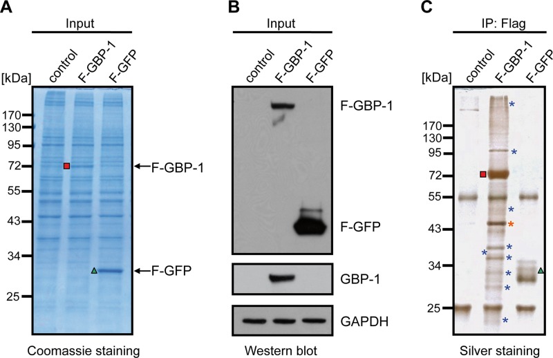
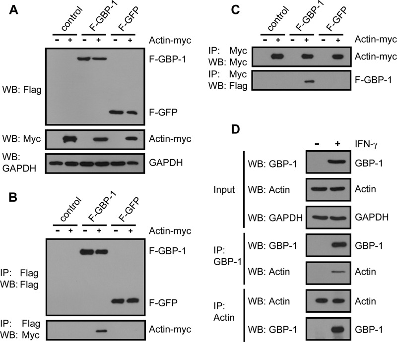
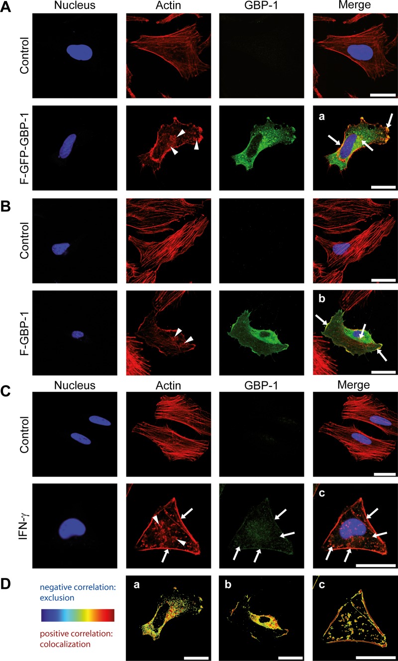
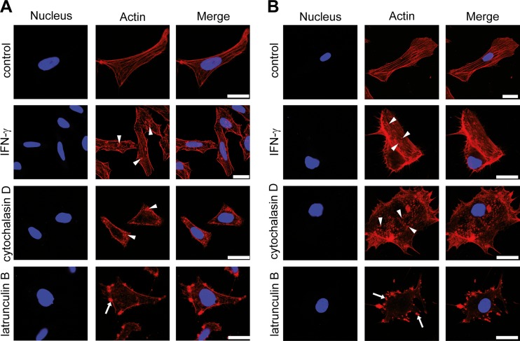
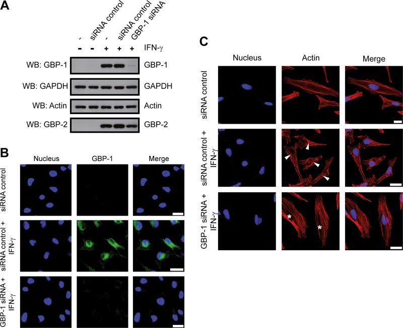
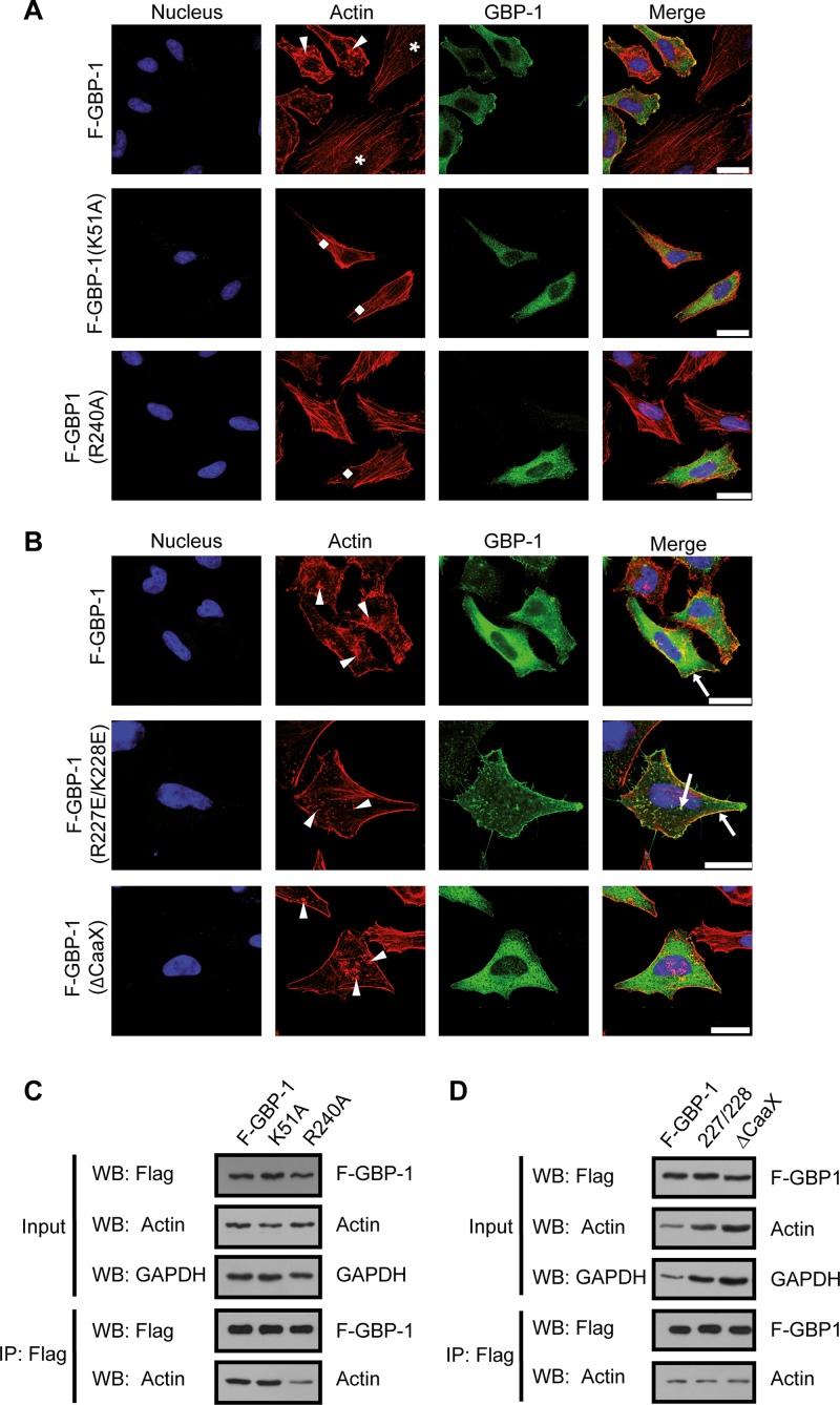
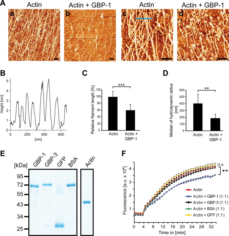
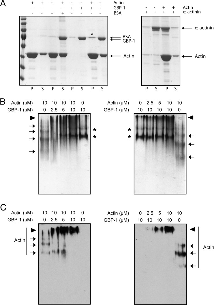
Gamma interferon (IFN-γ) regulates immune defenses against viruses, intracellular pathogens, and tumors by modulating cell proliferation, migration, invasion, and vesicle trafficking processes. The large GTPase guanylate binding protein 1 (GBP-1) is among the cellular proteins that is the most abundantly induced by IFN-γ and mediates its cell biologic effects. As yet, the molecular mechanisms of action of GBP-1 remain unknown. Applying an interaction proteomics approach, we identified actin as a strong and specific binding partner of GBP-1. Furthermore, GBP-1 colocalized with actin at the subcellular level and was both necessary and sufficient for the extensive remodeling of the fibrous actin structure observed in IFN-γ-exposed cells. These effects were dependent on the oligomerization and the GTPase activity of GBP-1. Purified GBP-1 and actin bound to each other, and this interaction was sufficient to impair the formation of actin filaments in vitro, as demonstrated by atomic force microscopy, dynamic light scattering, and fluorescence-monitored polymerization. Cosedimentation and band shift analyses demonstrated that GBP-1 binds robustly to globular actin and slightly to filamentous actin. This indicated that GBP-1 may induce actin remodeling via globular actin sequestering and/or filament capping. These results establish GBP-1 as a novel member within the family of actin-remodeling proteins specifically mediating IFN-γ-dependent defense strategies.


Figures

FIG 1
FIG 1
Identification of cellular interaction partners of human GBP-1 in HeLa cells. (A) HeLa cells were transfected with Flag-tagged GBP-1 (F–GBP-1) or with Flag-tagged GFP (F-GFP) or the empty parental vector (control) as controls. The protein lysates were separated via 10% SDS-PAGE and stained using Coomassie. Red square, GBP-1; green triangle, GFP. (B) Western blot analysis using antibodies against GBP-1 and the Flag tag. The detection of the GAPDH protein demonstrates equal protein loading. (C) Silver staining analysis of precipitates obtained via immunoprecipitation (IP) using an anti-Flag affinity gel with the various transfected cells, as described in the legend to panel A. Red square, F–GBP-1; green triangle, F-GFP; blue asterisks, potential interaction partners, which varied between the F–GBP-1 precipitate and the control precipitates; orange asterisk, actin.
FIG 2
FIG 2
Interaction of tagged and endogenous GBP-1 and actin proteins shown by reciprocal coimmunoprecipitation. (A) HeLa cells were cotransfected with F–GBP-1, F-GFP, or the empty parental vector each together with an expression plasmid encoding Myc-tagged actin (actin-Myc) or the corresponding control vector. Subsequently, 10 μg of lysate was analyzed via Western blotting (WB) using antibodies against the Flag and Myc tags. GAPDH was used as a loading control. (B) Immunoprecipitation analysis of the extracts from panel A with anti-Flag antibody and subsequent Western blot analysis with antibodies against either the Flag or Myc tag. (C) Lysates, transfected and analyzed as described in the legend to panel A, were immunoprecipitated using an antibody against the Myc tag. The precipitated proteins were subsequently analyzed via Western blotting using the indicated antibodies. (D) HeLa cells were either left untreated or treated with IFN-γ (100 U/ml, 24 h). The protein extracts were immunoprecipitated using an antibody against GBP-1 or actin and analyzed via Western blotting. GBP-1 and actin were detected using specific antibodies.
FIG 3
FIG 3
GBP-1 colocalizes with cellular actin and disrupts the stress fibers of the actin cytoskeleton. Images were taken using a Leica TCS SPE confocal microscope with z-plane focusing on the attachment site of the cell culture slide. Nuclei were counterstained with Draq5. (A) HeLa cells were transfected with an expression plasmid for Flag- and GFP-tagged GBP-1 (F–GFP–GBP-1) or the empty parental vector (control). F–GFP–GBP-1 was visualized by the fluorescence of the GFP tag, and actin was visualized using Alexa Fluor 546-labeled phalloidin. Arrowheads, the disrupted actin structure, observed as a granular distribution of actin proteins in the cytoplasm of F–GFP–GBP-1-transfected cells; arrows, colocalization of actin and F–GFP–GBP-1 observed at the plasma membrane and actin aggregates. (B) HUVECs were transfected with either F–GBP-1 or the empty parental vector (control). GBP-1 was detected in a subsequent immunofluorescence staining using an antibody against the Flag tag. Actin was detected by a fluorescently labeled phalloidin. The staining pattern was similar to that described in the legend to panel A. Arrows, colocalization. (C) HeLa cells were either left untreated (control) or treated with IFN-γ (100 U/ml, 24 h). Endogenous GBP-1 was stained using a GBP-1 antibody and immunofluorescence analysis. Arrowheads, impaired actin cytoskeleton shown as actin aggregates throughout the cytoplasm; arrows, colocalization of GBP-1 and actin. (D) The images from panels A to C (a, b, and c, respectively) were quantified using the ImageJ Colocalization Colormap software. The results are presented as images using hot colors (red and yellow), which display a positive correlation, and cold colors (blue), which display a negative correlation. A clear colocalization of GBP-1 and actin at the cell membrane, granular structures, and actin aggregates is shown. All results were independently reproduced by a second researcher. Bars = 25 μm.
FIG 4
FIG 4
The IFN-γ-induced disruption of the actin cytoskeleton mimics the effect of cytochalasin D. (A) HeLa cells were left untreated, treated with IFN-γ (100 U/ml, 24 h), or treated with different actin polymerization inhibitors (cytochalasin D and latrunculin B) (both at 0.2 μM, 30 min). Subsequently, the actin cytoskeleton was stained using fluorescently labeled phalloidin. Arrowheads, disrupted actin cytoskeleton, which was visualized as small, granular structures in the cytoplasm; arrows, bleb-like aggregates at the cell membrane in the latrunculin B-treated cells. The nuclei were counterstained with DAPI. (B) HUVECs were treated as described in the legend to panel A, with the exception that the polymerization inhibitor was utilized at a concentration of 0.1 μM. The actin pattern was observed as described in the legend to panel A and is indicated in the same manner described in the legend to panel A. The nuclei were counterstained with DAPI. Bars = 25 μm.
FIG 5
FIG 5
GBP-1 is necessary to mediate remodeling of the actin cytoskeleton induced by IFN-γ. (A) HeLa cells were transfected with either control siRNA or GBP-1 siRNA, as indicated. After 6 h, the cells were either left untreated or treated with IFN-γ (100 U/ml, 24 h). Next, 10 μg of protein lysate was separated via 10% SDS-PAGE and analyzed via Western blotting. GBP-1, actin, and GBP-2 (used as an siRNA specificity control) were detected using specific antibodies. (B) HeLa cells were transfected and treated with IFN-γ as described in the legend to panel A. The cells were stained for GBP-1 following antigen target retrieval (pH 9). The nuclei were counterstained with Draq5. (C) HeLa cells were treated as described in the legend to panel A and subsequently stained for actin. Asterisks, organized, filamentous actin cytoskeletons; arrowheads, disruption of the filamentous actin organization following IFN-γ treatment. The nuclei were counterstained with Draq5. All results were independently reproduced by a second researcher. Bars = 25 μm.
FIG 6
FIG 6
GBP-1 GTPase activity is required to induce remodeling of the actin cytoskeleton. (A) HeLa cells were transfected with expression constructs encoding F–GBP-1 and two GTPase-deficient mutant forms, F–GBP-1(R240A) and F–GBP-1(K51A). The immunocytochemical detection of GBP-1 was performed using an antibody against the Flag tag. Actin was visualized with fluorescently labeled phalloidin. Arrowheads, disrupted actin structures; lozenges, organized actin filaments. The nuclei are counterstained with Draq5. Note the different organizations of the cytoskeleton in F–GBP-1-expressing cells (arrowheads) and the nonexpressing cells (asterisks) in the upper panel. (B) F–GBP-1, a constitutively dimeric mutant [F–GBP-1(R227E/K228E)], and a mutant lacking the prenylation motif [F–GBP-1(ΔCaaX)] were overexpressed in HeLa cells. Immunofluorescence staining was performed as described in the legend to panel A. Arrowheads, a disrupted actin cytoskeleton structure; arrows, colocalization of GBP-1 and actin. The results shown in panels A and B were independently reproduced by a second researcher. (C) Flag immunoprecipitation and subsequent Western blot analysis of HeLa cells overexpressing proteins (as described in the legend to panel A). R240A, F–GBP-1(R240A); K51A, F–GBP-1(K51A). (D) Flag immunoprecipitation and Western blot analysis with HeLa cells overexpressing proteins (as described in the legend to panel B). 227/228, F–GBP-1(R227E/K228E); ΔCaaX, F–GBP-1(ΔCaaX). Bars = 25 μm.
FIG 7
FIG 7
GBP-1 directly interacts with actin in vitro. (A) Recombinant GBP-1, actin, gelsolin, and BSA proteins were detected via Western blotting using the indicated antibodies. (B) The recombinant proteins were mixed as indicated, and immunoprecipitations with either GBP-1 or actin were performed. The precipitates were analyzed via Western blotting using specific antibodies against GBP-1, BSA, gelsolin, or actin.
FIG 8
FIG 8
GBP-1 controls actin remodeling by reduction of filament length in vitro. (A) Actin (2 mg/ml) was polymerized alone or in the presence of GBP-1 (2 mg/ml). Polymerized actin filaments were deposited onto freshly cleaved, MgCl2-coated mica carriers and analyzed by AFM. Actin filaments were long in the absence of GBP-1 (a and c) and short in the presence of GBP-1 (b and d). White granular structures are due to salt precipitates. Bars = 400 nm. (B) Representative topography of actin filaments along the blue line (panel Ac) is given. Height measurements indicate an average diameter of actin filaments of 4 nm. (C) The relative actin filament length was quantitatively assessed by calculating the quotient of the total numbers of fibers and of the free polymer ends in five different optical fields. Actin fibers were significantly shorter in the presence of GBP-1 (***, P ≤ 0.001). (D) Actin (0.33 mg/ml) was polymerized in the presence or absence of GBP-1 (0.33 mg/ml), as described in the legend to panel A. Solutions were directly analyzed by DLS. For statistical analyses, the median of the hydrodynamic radius (D50) was averaged over six replicates per type. The standard deviation was calculated from the D50 values. **, P ≤ 0.01. (E) All purified proteins applied to the in vitro polymerization assays were separated by SDS-PAGE and visualized by Coomassie staining. (F) Actin polymerization using pyrene-labeled actin in combination with the indicated proteins was monitored over time by fluorescence intensity analysis. The polymerization assay was performed with pyrene-labeled actin alone or pyrene-labeled actin in combination with either GBP-1, GBP-3, BSA, or GFP. All proteins were applied at a 3 μM concentration. The results of one representative assay of three are shown. Significant differences between the experiments with GBP-1 and all other experimental conditions were observed after 5 min [ANOVA; F(7, 8) = 261; P ≤ 0.001]. GBP-1 showed a lower fluorescence than all other groups (P ≤ 0.008 each, HSD post hoc tests). a.u., arbitrary units. **, P < 0.01; n.s., not significant.
FIG 9
FIG 9
GBP-1 binds to both G-actin and F-actin. (A) F-actin was cosedimented by ultracentrifugation with GBP-1, BSA as a negative control, or α-actinin as a positive control. Equal amounts of pellets (lanes P) and supernatants (lanes S) were separated by SDS-PAGE. Gels were stained with Coomassie blue. An increase of GBP-1 in the pellet fraction was observed in the presence of F-actin, as indicated by an asterisk. (B) G-actin was incubated with GBP-1, and proteins were separated by native polyacrylamide gel electrophoresis. Gels were stained with Coomassie blue. Arrows, actin, which formed a ladder when incubated alone, indicating partial polymerization; asterisks, GBP-1 in its monomeric or dimeric form; arrowheads, actin–GBP-1 complexes. (C) Actin was detected using a specific antibody in a subsequent Western blot. Arrows, corresponding actin bands; arrowheads, larger protein complexes.

Similar articles

Cited by

References

    1. Damsker JM, Hansen AM, Caspi RR. 2010. Th1 and Th17 cells: adversaries and collaborators. Ann. N. Y. Acad. Sci. 1183:211–221. 10.1111/j.1749-6632.2009.05133.x - DOI - PMC - PubMed
    1. Ikeda H, Old LJ, Schreiber RD. 2002. The roles of IFN gamma in protection against tumor development and cancer immunoediting. Cytokine Growth Factor Rev. 13:95–109. 10.1016/S1359-6101(01)00038-7 - DOI - PubMed
    1. Bach EA, Aguet M, Schreiber RD. 1997. The IFN gamma receptor: a paradigm for cytokine receptor signaling. Annu. Rev. Immunol. 15:563–591. 10.1146/annurev.immunol.15.1.563 - DOI - PubMed
    1. Farrar MA, Schreiber RD. 1993. The molecular cell biology of interferon-gamma and its receptor. Annu. Rev. Immunol. 11:571–611. 10.1146/annurev.iy.11.040193.003035 - DOI - PubMed
    1. Stark GR, Kerr IM, Williams BR, Silverman RH, Schreiber RD. 1998. How cells respond to interferons. Annu. Rev. Biochem. 67:227–264. 10.1146/annurev.biochem.67.1.227 - DOI - PubMed

Publication types

LinkOut - more resources