. 2013 Nov 19;110(47):18964-9.
doi: 10.1073/pnas.1312783110. Epub 2013 Nov 4.

ADP-ribosyl-acceptor hydrolase 3 regulates poly (ADP-ribose) degradation and cell death during oxidative stress

Affiliations

ADP-ribosyl-acceptor hydrolase 3 regulates poly (ADP-ribose) degradation and cell death during oxidative stress

Masato Mashimo et al. Proc Natl Acad Sci U S A. .

Abstract

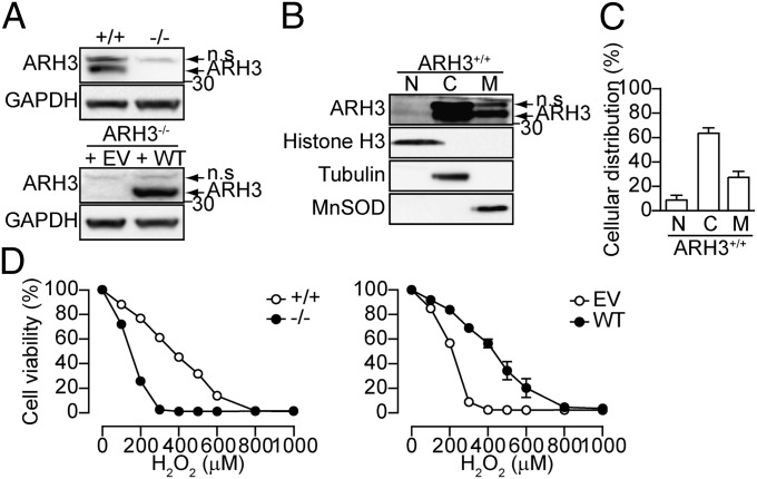
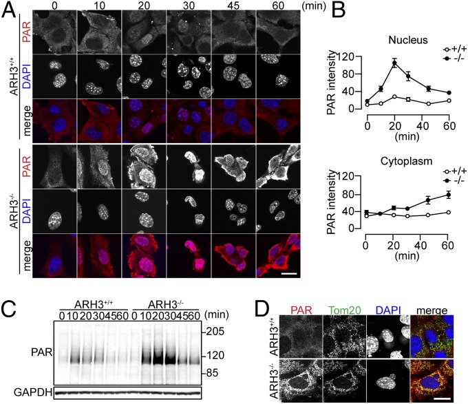
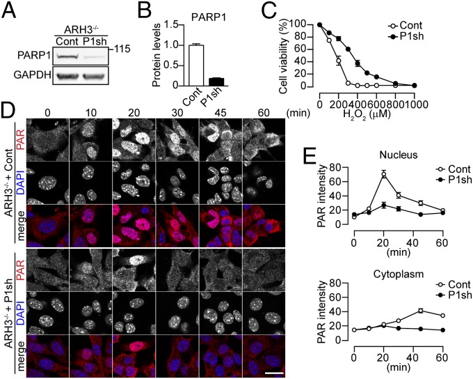
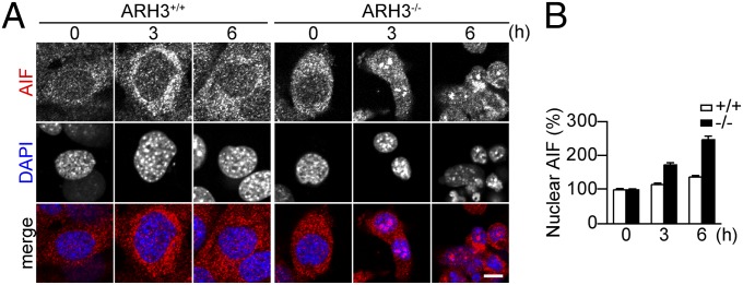
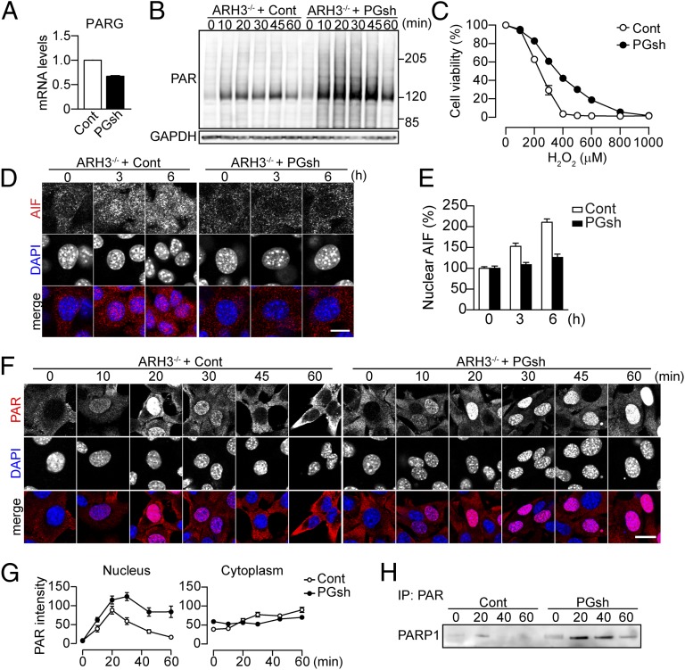
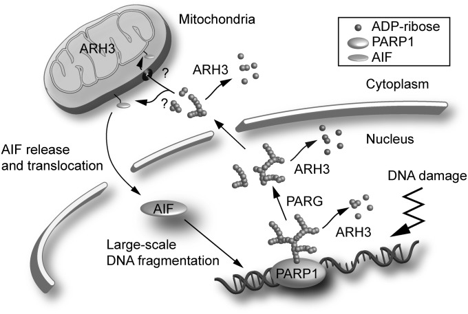
Poly (ADP ribose) (PAR) formation catalyzed by PAR polymerase 1 in response to genotoxic stress mediates cell death due to necrosis and apoptosis. PAR glycohydrolase (PARG) has been thought to be the only enzyme responsible for hydrolysis of PAR in vivo. However, we show an alternative PAR-degradation pathway, resulting from action of ADP ribosyl-acceptor hydrolase (ARH) 3. PARG and ARH3, acting in tandem, regulate nuclear and cytoplasmic PAR degradation following hydrogen peroxide (H2O2) exposure. PAR is responsible for induction of parthanatos, a mechanism for caspase-independent cell death, triggered by apoptosis-inducing factor (AIF) release from mitochondria and its translocation to the nucleus, where it initiates DNA cleavage. PARG, by generating protein-free PAR from poly-ADP ribosylated protein, makes PAR translocation possible. A protective effect of ARH3 results from its lowering of PAR levels in the nucleus and the cytoplasm, thereby preventing release of AIF from mitochondria and its accumulation in the nucleus. Thus, PARG release of PAR attached to nuclear proteins, followed by ARH3 cleavage of PAR, is essential in regulating PAR-dependent AIF release from mitochondria and parthanatos.

Keywords: cytotoxicity; posttranslational modification.


Conflict of interest statement

The authors declare no conflict of interest.

Figures

Fig. 1.
Fig. 1.
ARH3 protected against H2O2-induced cell death. (A) ARH3 expression. Cells were subjected to Western blotting by using anti-ARH3 antibody. GAPDH was used as a loading control. Note that an upper band reactive with anti-ARH3 antibody is nonspecific (n.s.). (B) Intracellular ARH3 distribution. Identity of fractions without cross-contamination was confirmed by localization of histone H3 (nucleus), tubulin, (cytoplasm), and manganese superoxide dismutase (MnSOD, mitochondria). (C) Quantification of intracellular ARH3 distribution (means ± SEM, n = 7). (D) H2O2-induced cell death. Cells were exposed to H2O2 (24 h) at indicated concentrations before assessment of cell viability (means ± SEM, n = 3). These representative data (A and B) have been replicated three times with similar results.
Fig. 2.
Fig. 2.
ARH3 regulated nuclear and cytoplasmic PAR content in response to H2O2. (A) Time-dependent PAR localization after 300 μM H2O2 exposure for indicated times. Cells were subjected to immunocytochemistry by using anti-PAR antibody (red in merged images) and DAPI staining (blue in merged images). (B) Mean PAR fluorescence in nuclei and cytoplasm (means ± SEM, n = 6–40 cells). (C) Time course of H2O2-induced PAR accumulation after 300 μM H2O2 exposure for indicated times. Cells were subjected to Western blotting by using anti-PAR antibody. GAPDH was used as a loading control. (D) PAR localization after 2-h exposure to 300 μM H2O2. Cells were subjected to immunocytochemistry by using anti-PAR (red in merged images) and Tom20 antibodies (green in merged images) and DAPI staining (blue in merged images). These representative data (A, C, and D) have been replicated three times with similar results. (Scale bars: 20 μm.)
Fig. 3.
Fig. 3.
PARP1-mediated PAR synthesis and cell death in response to H2O2 in ARH3−/− MEFs. (A) PARP1 expression. Cells were subjected to Western blotting by using anti-PARP1 antibody. (B) Quantification of PARP1 expression levels. The amount of PARP1 protein was normalized to that of GAPDH (means ± SEM, n = 3). (C) Effect of PARP1 depletion on ARH3−/− MEFs viability after H2O2 exposure. Cells were exposed to H2O2 (24 h) at indicated concentrations before assessment of cell viability (means ± SEM, n = 3). (D) Time-dependent PAR localization after 300 μM H2O2 exposure. Cells were subjected to immunocytochemistry by using anti-PAR antibody (red in merged images) and DAPI staining (blue in merged images). (Scale bar: 20 μm.) (E) Mean PAR fluorescence in nuclei and cytoplasm (means ± SEM, n = 39–40 cells). These representative data (A and D) have been replicated three times with similar results.
Fig. 4.
Fig. 4.
ARH3 deficiency enhanced AIF accumulation in the nucleus after H2O2 exposure. (A) AIF accumulation in nuclei of ARH3−/− MEFs after 3- or 6-h exposure to 300 μM H2O2. Cells were subjected to immunocytochemistry by using anti-AIF antibody (red in merged images) and DAPI staining (blue in merged images). (Scale bar: 10 μm.) These representative data have been replicated three times with similar results. (B) Mean AIF fluorescence in nuclei (means ± SEM, n = 36–40 cells).
Fig. 5.
Fig. 5.
Partial depletion of PARG enhanced PAR accumulation and decreased AIF-mediated cell death in response to H2O2 in ARH3−/− MEFs. (A) Effect of shRNA on PARG mRNA. PARG mRNA was normalized to GAPDH mRNA (means ± SEM, n = 3). (B) Time course of H2O2-induced PAR accumulation after exposure to 300 μM H2O2 for indicated times. Cells were subjected to Western blotting by using anti-PAR antibody. GAPDH was used as a loading control. (C) Effect of PARG depletion on ARH3−/− MEF viability after H2O2 exposure. Cells were exposed to H2O2 (24 h) at indicated concentrations before assessment of cell viability (means ± SEM, n = 3). (D) AIF accumulation in nuclei of ARH3−/− MEFs after 3- or 6-h exposure to 300 μM H2O2. Cells were subjected to immunocytochemistry by using anti-AIF antibody (red in merged images) and DAPI staining (blue in merged images). (E) Mean AIF fluorescence in nuclei (means ± SEM, n = 40 cells). (F) Time-dependent PAR localization after 300 μM H2O2 exposure for indicated times. Cells were subjected to immunocytochemistry by using anti-PAR antibody (red in merged images) and DAPI staining (blue in merged images). (G) Mean PAR fluorescence in nuclei and cytoplasm (means ± SEM, n = 38–40 cells). (H) Poly-ADP ribosylated PARP1 after 300 μM H2O2 exposure for indicated times. Cells were subjected to immunoprecipitation by using anti-PAR antibody and then Western blotting by using anti-PARP1 antibody to detect poly-ADP ribosylated PARP1. These representative data (B, D, F, and H) have been replicated three times with similar results. (Scale bars: D, 10 μm; F, 20 μm.)
Fig. 6.
Fig. 6.
Model for a role of ARH3 in PAR degradation and AIF-mediated cell death. Overactivation of PARP1 by widespread DNA damage results in poly-ADP ribosylation of PARP1 and other acceptor proteins in the nucleus. PARG hydrolyzes PAR attached to acceptor proteins such as PARP1 to generate protein-free small PAR molecules, thereby facilitating its translocation to the cytoplasm and mitochondria. ARH3 located in the nucleus and cytoplasm hydrolyzes PAR. PAR may bind to AIF anchored in mitochondrial membrane, releasing it to the cytoplasm. Once in the cytoplasm, AIF translocates to the nucleus via its nuclear localization signal. In nucleus, AIF recruits nucleases such as cyclophilin A and H2AX, resulting in large-scale DNA fragmentation. ARH3 is also located in the mitochondrial matrix.

References

    1. Schreiber V, Dantzer F, Ame JC, de Murcia G. Poly(ADP-ribose): Novel functions for an old molecule. Nat Rev Mol Cell Biol. 2006;7(7):517–528. - PubMed
    1. Amé JC, Spenlehauer C, de Murcia G. The PARP superfamily. Bioessays. 2004;26(8):882–893. - PubMed
    1. Ueda K, Hayaishi O. ADP-ribosylation. Annu Rev Biochem. 1985;54:73–100. - PubMed
    1. Masson M, et al. XRCC1 is specifically associated with poly(ADP-ribose) polymerase and negatively regulates its activity following DNA damage. Mol Cell Biol. 1998;18(6):3563–3571. - PMC - PubMed
    1. D’Amours D, Desnoyers S, D’Silva I, Poirier GG. Poly(ADP-ribosyl)ation reactions in the regulation of nuclear functions. Biochem J. 1999;342(Pt 2):249–268. - PMC - PubMed

Publication types

Substances