

Modulation of murine macrophage TLR7/8-mediated cytokine expression by mesenchymal stem cell-conditioned medium
- PMID: 24191131
- PMCID: PMC3804401
- DOI: 10.1155/2013/264260
Modulation of murine macrophage TLR7/8-mediated cytokine expression by mesenchymal stem cell-conditioned medium
Abstract
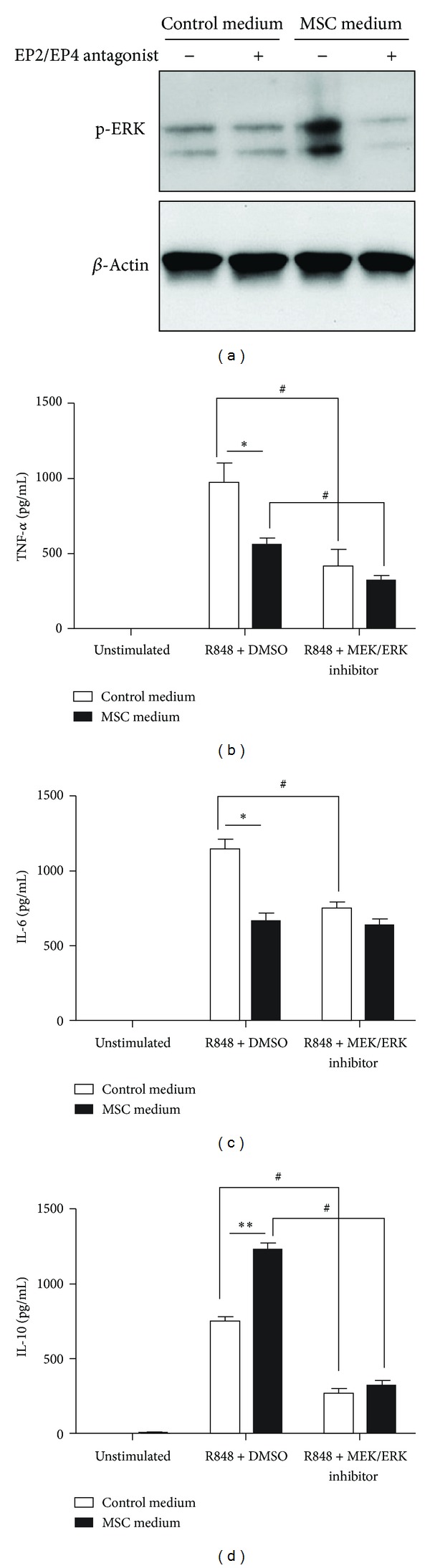
Increasing evidence suggests that mesenchymal stem cells (MSCs) play anti-inflammatory roles during innate immune responses. However, little is known about the effect of MSCs or their secretions on the ligand response of Toll-like receptor (TLR) 7 and TLR8, receptors that recognize viral single-stranded RNA (ssRNA). Macrophages play a critical role in the innate immune response to ssRNA virus infection; therefore, we investigated the effect of MSC-conditioned medium on cytokine expression in macrophages following stimulation with TLR7/8 ligands. After stimulation with TLR7/8 ligand, bone marrow-derived macrophages cultured with MSCs or in MSC-conditioned medium expressed lower levels of tumor necrosis factor (TNF) α and interleukin (IL) 6 and higher levels of IL-10 compared to macrophages cultured without MSCs or in control medium, respectively. The modulations of cytokine expression were associated with prostaglandin E2 (PGE2) secreted by the MSCs. PGE2 enhanced extracellular signal-related kinase (ERK) signaling and suppressed nuclear factor- κ B (NF- κ B) signaling. Enhanced ERK signaling contributed to enhanced IL-10 production, and suppression of NF- κ B signaling contributed to the low production of TNF- α . Collectively, these results indicate that MSCs and MSC-conditioned medium modulate the cytokine expression profile in macrophages following TLR7/8-mediated stimulation, which suggests that MSCs play an immunomodulatory role during ssRNA virus infection.
Figures


Similar articles
-
Sensitivity of TLR4- and -7-induced NF kappa B1 p105-TPL2-ERK pathway to TNF-receptor-associated-factor-6 revealed by RNAi in mouse macrophages.Mol Immunol. 2007 Jul;44(15):3715-23. doi: 10.1016/j.molimm.2007.04.002. Epub 2007 May 15. Mol Immunol. 2007. PMID: 17507094
-
Differential TLR-ERK1/2 Activity Promotes Viral ssRNA and dsRNA Mimic-Induced Dysregulated Immunity in Macrophages.Pathogens. 2024 Nov 23;13(12):1033. doi: 10.3390/pathogens13121033. Pathogens. 2024. PMID: 39770293 Free PMC article.
-
TLR7 Ligation Inhibits TLR8 Responsiveness in IL-27-Primed Human THP-1 Monocytes and Macrophages.J Innate Immun. 2021;13(6):345-358. doi: 10.1159/000515738. Epub 2021 May 31. J Innate Immun. 2021. PMID: 34058746 Free PMC article.
-
The Multifunction Role of Tumor-Associated Mesenchymal Stem Cells and Their Interaction with Immune Cells in Breast Cancer.Immunol Invest. 2023 Nov;52(7):856-878. doi: 10.1080/08820139.2023.2249025. Epub 2023 Aug 24. Immunol Invest. 2023. PMID: 37615117 Review.
-
Role of toll-like receptors in modulation of cytokine storm signaling in SARS-CoV-2-induced COVID-19.J Med Virol. 2022 Mar;94(3):869-877. doi: 10.1002/jmv.27405. Epub 2021 Oct 26. J Med Virol. 2022. PMID: 34672376 Free PMC article. Review.
Cited by
-
Effect of Placenta-Derived Mesenchymal Stromal Cells Conditioned Media on an LPS-Induced Mouse Model of Preeclampsia.Int J Mol Sci. 2022 Jan 31;23(3):1674. doi: 10.3390/ijms23031674. Int J Mol Sci. 2022. PMID: 35163594 Free PMC article.
-
Mesenchymal Stem Cell-Macrophage Choreography Supporting Spinal Cord Repair.Neurotherapeutics. 2018 Jul;15(3):578-587. doi: 10.1007/s13311-018-0629-0. Neurotherapeutics. 2018. PMID: 29728851 Free PMC article. Review.
-
Role of mesenchymal stem cell derived extracellular vesicles in autoimmunity: A systematic review.World J Stem Cells. 2020 Aug 26;12(8):879-896. doi: 10.4252/wjsc.v12.i8.879. World J Stem Cells. 2020. PMID: 32952864 Free PMC article.
-
Cerebrospinal Fluid (CSF) Exchange with Artificial CSF Enriched with Mesenchymal Stem Cell Secretions Ameliorates Experimental Autoimmune Encephalomyelitis.Int J Mol Sci. 2019 Apr 11;20(7):1793. doi: 10.3390/ijms20071793. Int J Mol Sci. 2019. PMID: 30978957 Free PMC article.
-
Regenerative medicine using dental pulp stem cells for liver diseases.World J Gastrointest Pharmacol Ther. 2017 Feb 6;8(1):1-6. doi: 10.4292/wjgpt.v8.i1.1. World J Gastrointest Pharmacol Ther. 2017. PMID: 28217369 Free PMC article.
References
-
- Prendergast AJ, Klenerman P, Goulder PJ. The impact of differential antiviral immunity in children and adults. Nature Reviews Immunology. 2012;12(9):636–648. - PubMed
-
- Kawai T, Akira S. Antiviral signaling through pattern recognition receptors. Journal of Biochemistry. 2007;141(2):137–145. - PubMed
-
- Wu S, Metcalf JP, Wu W. Innate immune response to influenza virus. Current Opinion in Infectious Diseases. 2011;24(3):235–240. - PubMed
Publication types
MeSH terms
Substances
LinkOut - more resources
Full Text Sources
Other Literature Sources
Miscellaneous
