Arthroscopic anatomy of the subdeltoid space
- PMID: 24191185
- PMCID: PMC3808800
- DOI: 10.4081/or.2013.e25
Arthroscopic anatomy of the subdeltoid space
Abstract
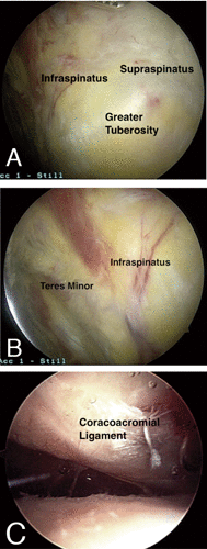
From the first shoulder arthroscopy performed on a cadaver in 1931, shoulder arthroscopy has grown tremendously in its ability to diagnose and treat pathologic conditions about the shoulder. Despite improvements in arthroscopic techniques and instrumentation, it is only recently that arthroscopists have begun to explore precise anatomical structures within the subdeltoid space. By way of a thorough bursectomy of the subdeltoid region, meticulous hemostasis, and the reciprocal use of posterior and lateral viewing portals, one can identify a myriad of pertinent ligamentous, musculotendinous, osseous, and neurovascular structures. For the purposes of this review, the subdeltoid space has been compartmentalized into lateral, medial, anterior, and posterior regions. Being able to identify pertinent structures in the subdeltoid space will provide shoulder arthroscopists with the requisite foundation in core anatomy that will be required for challenging procedures such as arthroscopic subscapularis mobilization and repair, biceps tenodesis, subcoracoid decompression, suprascapular nerve decompression, quadrangular space decompression and repair of massive rotator cuff tears.
Keywords: anatomy; arthroscopy; subdeltoid space.
Conflict of interest statement
Conflict of interests: the authors declare no potential conflict of interests.
Figures


Similar articles
-
All-Arthroscopic Modified Rotator Interval Slide for Massive Anterosuperior Cuff Tears Using the Subdeltoid Space: Surgical Technique and Early Results.HSS J. 2016 Oct;12(3):200-208. doi: 10.1007/s11420-016-9497-5. Epub 2016 Mar 24. HSS J. 2016. PMID: 27703412 Free PMC article.
-
[Massive tears of the rotator cuff--comparison of mini-open and arthroscopic techniques. Part 2. Arthroscopic repair].Acta Chir Orthop Traumatol Cech. 2007 Oct;74(5):318-25. Acta Chir Orthop Traumatol Cech. 2007. PMID: 18001628 Czech.
-
The arthroscopic "subdeltoid approach" to the anterior shoulder.J Shoulder Elbow Surg. 2013 Apr;22(4):e6-10. doi: 10.1016/j.jse.2012.09.006. Epub 2013 Jan 11. J Shoulder Elbow Surg. 2013. PMID: 23313368
-
Subscapularis tears: hidden and forgotten no more.JSES Open Access. 2018 Mar 1;2(1):74-83. doi: 10.1016/j.jses.2017.11.006. eCollection 2018 Mar. JSES Open Access. 2018. PMID: 30675571 Free PMC article. Review.
-
Arthroscopic management of rotator cuff disease.J Am Acad Orthop Surg. 1998 Jul-Aug;6(4):259-66. doi: 10.5435/00124635-199807000-00007. J Am Acad Orthop Surg. 1998. PMID: 9682088 Review.
Cited by
-
Reliability of a simple physical therapist screening tool to assess errors during resistance exercises for musculoskeletal pain.Biomed Res Int. 2014;2014:961748. doi: 10.1155/2014/961748. Epub 2014 Mar 13. Biomed Res Int. 2014. PMID: 24738079 Free PMC article.
-
Effect of video-based versus personalized instruction on errors during elastic tubing exercises for musculoskeletal pain: a randomized controlled trial.Biomed Res Int. 2014;2014:790937. doi: 10.1155/2014/790937. Epub 2014 Mar 10. Biomed Res Int. 2014. PMID: 24734244 Free PMC article. Clinical Trial.
References
-
- Ruotolo C, Fow JE, Nottage WM. The supraspinatus footprint: an anatomic study of the supraspinatus insertion. Arthroscopy 2004;20:246-9 - PubMed
-
- Mochizuki T, Sugaya H, Uomizu M, et al. Humeral insertion of the supraspinatus and infraspinatus. New anatomical findings regarding the footprint of the rotator cuff J Bone Joint Surg Am 2008;90:962-9 - PubMed
-
- Nicholson GP, Goodman DA, Flatow EL, Bigliani LU. The acromion: morphologic condition and age-related changes. A study of 420 scapulas J Shoulder Elbow Surg 1996;5:1-11 - PubMed
-
- Aoki M IS, Usui M. The slope of the acromion in the rotator cuff impingement. Orthop Trans 1986;10:228
-
- Holt EM, Allibone RO. Anatomic variants of the coracoacromial ligament. J Shoulder Elbow Surg 1995;4:370-5 - PubMed
Publication types
LinkOut - more resources
Full Text Sources
Other Literature Sources
